Download
1 / 82
820 likes | 1.01k Views
Cardiovascular Emergencies Part 1. Assessment. Primary survey/ resuscitation Secondary survey. Unmodifiable Age Sex Heredity Race. Modifiable B/P Obesity Dyslipidemia Smoking Sedentary life style Stress Diabetes. Factors For Consideration. Focused Survey. Subjective data

E N D
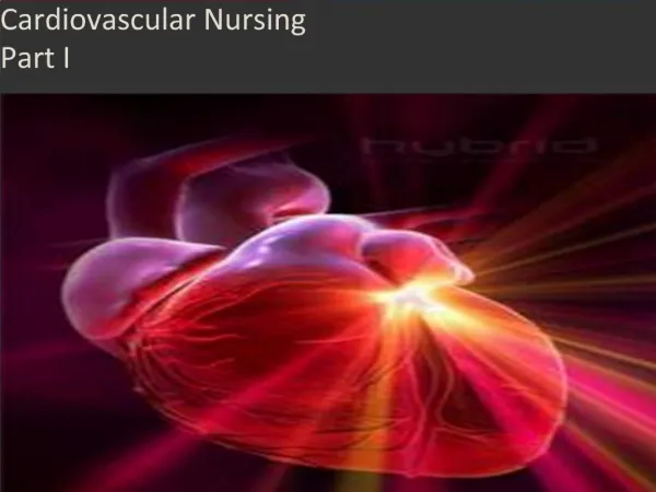
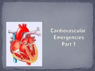
Cardiovascular Emergencies Part 1
Assessment • Primary survey/ • resuscitation • Secondary survey
Unmodifiable Age Sex Heredity Race Modifiable B/P Obesity Dyslipidemia Smoking Sedentary life style Stress Diabetes Factors For Consideration
Focused Survey Subjective data • Chief complaint • History of present illness • Pain: PQRST • Provocation • Quality • Region/radiation • Severity • Time • Duration • Development over time • Periodicity (? Comes/goes)
Dyspnea SOB Dyspnea on exertion Positional dyspnea Paroxysmal nocturnal dyspnea Orthopnea Cough Dry, “cardiac” cough Hemoptysis Syncope Palpitations Fatigue Nausea and Vomiting Headache Behavioral change Activity limitations Injury: mechanism and time Focused Survey (Continued)
Medical History • Coronary Heart Disease • Angina • Previous MI • Hypertension • CHF • Pulmonary Disease • Diabetes • Renal Disease • Previous Cardiac Surgery • Congenital Anomalies • Allergies
Current/Past Medication Use • Nitrates • Beta Blockers • Calcium Channel Blockers • Anti- hypertensives • Digitalis • Diuretics • Antidysrhythmics • Anticoagulants • Steroids • Specific Pulmonary Drugs • Illicit Drugs • OVC Medications
Physical Exam • General Survey • LOC • Respiratory status • Rate, regularity, effort, breath sounds • Integumentary • Color, temperature, moisture, capillary refill • Edema • Dependent, extremities, sacrum, pleural effusion, ascitis, cardiac (pitting)
Physical Exam 0 = absent 1 = thready 2 = normal 3 = bounding • Cyanosis • Central • Peripheral • Clubbing • B/P Measurement • Both arms • Orthostatic (supine, sitting, standing)
Physical Exam • Apical heart rate • Regular, irregular, regularly irregular, irregular irregular • Peripheral pulses • Pupils • Size, equality, reactive
Inspection • Tracheal position • Neck veins • Can guesstimate CVP • Thorax • Configuration • Deformities, anterior, posterior, A-P diameter, symmetrical movement • Injuries, penetrating, blunt (ecchymosis, contusions) evidence of scars, surgery • Abnormal chest movement, asymmetrical, paradoxical
Physical Exam (Inspection) • Precordium • Apical pulse • Abnormal precordial movements, heaves, lifts, pulsations, retractions • Epigastrium, pulsations
Physical Exam: Palpation • Areas tender, or crepitus • Epigastrium
Auscultatorysites Murmurs: systolic/diastolic Variations in rhythm Extra sounds Pericardial friction rub Venous hum Arterial bruits Clicks Physical Exam: Auscultation
Physical Exam: Diagnostic Procedures • ECG, 12 lead • Rate, rhythm • Presence of cardiac dysrhythmias • Evidence of myocardial ischemia, injury • Presence of intraventricular conduction defect • Evidence of previous MI
Physical Exam:Diagnostic Procedures • CBC • Serum electrolytes • Cardiac serum markers • Troponin • Myoglobin • Basic metabolic profile • Coagulation studies • Digoxin level • Serum creatinine, BUN • T& C • ABGs • Routine urinalysis
Pulse oximetry Radiography Chest x-ray Heart size and location Presence of edema Pulmonary infiltrates Pleural effusions Air and fluid levels in trauma patients Mediastinal width Bony structure integrity Cardiac catheterization Echocardiogram Diagnostic Procedures
Interventions • Determine priorities • Airway, vital signs, cardiac rhythm, ABGs, Pulse oximetry, • Control pain • Relieve anxiety • Education patient/other • Prevent complications • Establish care plan • Emergency equipment • Initiate appropriate interventions • Document data • Monitor responses and adjust
Pediatrics • Growth & Development • R/T congenital heart disease (heart defects) • Acquired heart disease (rheumatic fever) • Endocrine (diabetes) • Other • Drug ingestions, ex: tricyclics, digoxin • Trauma (falls, MVCs) • Suffocation (plastic bags, drowning, accidental hanging)
Pediatric • Pearls: • Cardiac arrest usually d/t progressive deterioration in respiratory and heart function • CHF, cardiogenic shock, dysrhythmias are unusual. If occur congenital. • Immature conduction system and autonomic innervation may contribute to dysrhythmias
Presence of chronic diseases Altered drug metabolism Multiple physiological differences and changes in lab values must be considered when assessing the older patient Psychological and social changes: patient may have different goals for their treatment, discuss with patient Geriatric patients can adapt to disease so well that symptoms are not obvious Arteriosclerotic changes in aorta and peripheral pulses may pose a difficulty in palpating Rhythm abnormalities are so common that they may be “normal” Geriatric
Pearls • “Go slow, stay low” with medications • Concurrent use of other medications cause problems, • Easy to use meds are helpful (transdermals) • One time or two time doses daily • Evaluate medications on a frequent basis
Angina • Stable • Symptom of ischemia • “pain or discomfort” • Poorly localized • Flow/Demand imbalance • May be chronic, acute, or unstable
Angina (continued) • Unstable angina • New symptoms of angina • Increasing symptoms that occur at rest or with on exertion • Usually due to platelet aggregation • Leads to atypical chest pain
Angina (Continued) • Unstable angina diagnosis • Angina is at rest, as well as minimal exertion (usually 20 minutes or longer) • Angina of new onset (several weeks), starting with physical exertion, and markedly limits activity • Previously diagnosed stable angina
Angina cont’d • Variant Angina • May or may not be due to atherotic changes • Thought to be due to coronary spasm • Prinzmetal’sangina • May occur at the same time, daily • Usually not associated with exertion or stress • Occurs at younger ages • ST elevation seen during pain, then disappears
Angina: Curveballs • GERDs • Biliary Colic • Chest Wall Pain • Pericarditis • PE • Aortic Dissection • Dysrhythmias
Angina: Data • Physical exam: • Elevated B/P • Increased Heart Rate • Rapid, shallow respirations • Diaphoresis • Transient pulmonary rales (with pain)
Diagnostic Procedures: ECG • ST depression may accompany pain with stable angina • Transient ST-segment deviations (depression or elevation), and T wave inversion occur commonly with unstable angina • Variant angina: ST elevation occurs with pain, subsides when pain does • May see LV hypertrophy, old MI, nonspecific ST and T-wave abnormalities and AV defects
Diagnostic Procedures (Continued) • CBC • Cardiac serum markers…no elevation should occur unless cell damage • Chest X-ray ( CHF, cardiomegaly)
Interventions • Continuous monitoring • O2 • IV, Draw labs • 12 Lead ECG • Rest • Decrease anxiety • SL NTG, B/P 100 mm HG followed by a drip • Assess for H/A • Reflex tachycardia • Cautious with elderly
Interventions (continued) • Beta blockers • If clinical situation deteriorates after B Blocker, consider coronary artery spasm • Assess for signs of heart failure • Adverse effects of blockers are considered more common and severe in geriatric
Interventions (continued) • Administer antiplatelet agents • ASA (4-5 baby aspirin) • Administer ASAP • Decreases platelet activation and thrombus formation • TEACH/EDUCATION
MI: Data • Physical exam • General appearance: Anxious, restless, clenched fist against chest (Levine sign) Look of doom • Heart rate: may be ok, tachycardia (most common), bradycardia (inferior and RV), Regular or irregular PVCs common
Data (continued) • Arterial BP • Majority of patients with uncomplicated MI are normotensive • May be elevated due to SNS stimulation • Pain and anxiety • Decreased as a result of impaired cardiac function or due to drug administration (nitrates, M.S.)
Data (continued) • Respiratory rate: Initially elevated. Should return to normal after pain relief • Patients with CHF, respiratory rate correlates with severity of condition
Data (continued) • Peripheral: How bad is the patient’s condition? • Pallor, cyanosis, diaphoreses, mottled, cool, peripheral pulses variable • Temperature: Often increases 4-8 hours post MI • Heart Sounds: muffled. murmurs may be transient or permanent
Diagnosis • Continuous cardiac monitoring • 12 lead ECG • Determine Location of infarct (next slide)