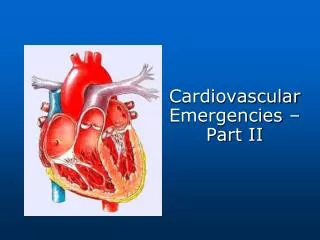
Cardiovascular Emergencies – Part II
760 likes | 1.1k Views
Cardiovascular Emergencies – Part II. Acute Aortic Dissection. Uncommon but lethal! Tear in the intimal layer of the aorta that results in a false lumen that is usually anterograde in nature. Usual locations: ascending aorta superior to aortic valve

Cardiovascular Emergencies – Part II
E N D
Presentation Transcript
Acute Aortic Dissection • Uncommon but lethal! • Tear in the intimal layer of the aorta that results in a false lumen that is usually anterograde in nature. • Usual locations: • ascending aorta superior to aortic valve • descending aorta at the ligamentum arteriosm
Acute Aortic Dissection The most common and most lethal acute aortic dissection, which accounts for 2/3 of all dissections, occurs where?
Most common in men between the ages of 60 & 70 Factors: hypertension hereditary defects of connective tissue (Marfan’s) pregnancy blunt trauma iatrogenic factors (intra-arterial catheterization) Acute Aortic Dissection
Acute Aortic Dissection SUBJECTIVE DATA • History • Pain – sudden, sharp, tearing, excruciating, medications may not relieve, substernal (ascending), back/flank (descending) • Syncope • Altered LOC • Paraplegia
Acute Aortic Dissection OBJECTIVE DATA • Physical Exam - variable BPs on right vs left - decreased peripheral pulses/ peripheral cyanosis - murmur - pallor, oliguria, altered LOC, - BP: hyper with distal dissection, hypo with proximal - extreme pain
Acute Aortic Dissection OBJECTIVE DATA • Diagnostics - CBC (Hct tends to fall, WBC 12,000-20,000) T&C,BUN/Creatinine - EKG: Normal in 1/3, LV hypertrophy if hx of HTN, signs of MI if proximal dissection -
Acute Aortic Dissection CXR: -widened aortic silhouette -widened mediastinum, -left-sided pleural effusion
Acute Aortic Dissection • Diagnostics cont. - CT Scan
Acute Aortic Dissection INTERVENTIONS • ABC • Pain relief • Large bore IVs • – minimum of two sites • Monitoring • Medications: • to lower arterial BP: nitroprusside, labetalol
Acute Aortic Dissection • Medications cont: 2) To decrease contraction force: beta blockers preferred, may give calcium channel blockers if beta blockers contraindicated 3) To relieve pain: Morphine • Position of comfort • IVF in hypotensive setting • Foley
Acute Aortic Dissection • Anticipate: ED thoracotomy, immediate need for OR, arterial & central venous cannulation • Therapeutics: Explain all procedures to patient/family, maintain calm, allow family at bedside if possible
Acute Pericarditis • Result of inflammation of the pericardium that may extend to adjacent structures and may produce exudate. • Factors: - infections: idiopathic, viral, bacterial, fungal - connective tissue disease (lupus, rheumatoid) - renal disease - neoplastic disorders - tissue injury
Acute Pericarditis Acute pericarditis is more common in which gender and which age group?
Acute Pericarditis SUBJECTIVE DATA • General malaise, fever, chills, weight loss • Dyspnea, cough • Chest Pain – deep inspiration, recumbent, movement, severe, sharp or dull ache, retrosternal or epigastric radiating to back/neck/ side, sudden, persistent
Acute Pericarditis SUBJECTIVE DATA cont. • Medical History may include: • TB, congenital anomalies, immune disorders, MI, neoplastic disease, drug use, uremia, cardiac surgery, cardiac trauma, infections
Acute Pericarditis OBJECTIVE DATA • Physical Exam - pericardial friction rub (hallmark) – heard best at the left lower sternum during end expiration with patient leaning forward - tachycardia, fever, tachypnea
Acute Pericarditis Diagnostics - EKG: FOUR STAGES (best seen in inferior leads) 1) ST elevation (early) with upright T waves, 2) T wave flattens and ST returns to baseline 3) T wave inversion 4) T wave returns to normal (weeks to months) - Lab: CBC, BUN/Cr, Electrolytes, cultures, UA - CXR: helpful to detect pericardial effusion or potential etiology - Echocardiogram: most accurate in detecting!!
Acute Pericarditis INTERVENTIONS • Supplemental O2, cardiac monitoring • Position of comfort • Anti-inflammatory medications • Pericardiocentesisif necessary • Labs as ordered • Antibiotics as ordered
Acute Pericarditis INTERVENTIONS cont • Monitor/reassess • Therapeutics: • maintain calm • explain all procedures • allow family at bedside if possible • reassurance
Infective Endocarditis Infection of the endocardium and heart valves • SBE • subacute bacterial endocarditis usually occurs in patients with congenital or acquired valvular disease; patients are less toxic • ABE • acute bacterial endocariditis usually affects normal heart valves and has a greatly accelerated pace of development; patients are extremely toxic with metastatic infections.
Infective Endocarditis • Infective agents (most common): - ABE: staphylococcus aureus - SBE: streptcoccus viridans • Risk factors: - Valvular disease, congenital heart defects, rheumatic heart disease, prosthetic heart valves, IV drug abusers, LT vascular access catheters
Infective Endocarditis • General pathophysiology: • platelets and fibrin deposit on abnormal endothelium • organisms adhere and colonization begins • microorganisms or fragments shed into blood • infarction or infection can occur at any distal site • infection of cardiac tissue can lead to progressive heart failure, conduction disturbances, and dysrhythmias.
Infective Endocarditis Which age population is infective endocariditis rarely seen in?
Infective Endocarditis SUBJECTIVE DATA • Fever: SBE – low grade, ABE – 102 degrees F • Anorexia, weight loss, night sweats • Arthralgia, myalgia, fatigue, malaise • Dyspnea, cough, pleuritic chest pain, hemoptysis • HA, signs of stroke, confusion • Abdominal and back pain
Cardiac surgery Congenital or aquired heart valve disease IV drug use Rheumatic heart disease Cardiac pacemaker Recent GI or GU disorder with valve disease Prosthetic valves with recent dental procedures without prophylactic ATX Infective Endocarditis Subjective Data Suspect if history of:
Infective Endocarditis OBJECTIVE DATA • Fever – may be absent in elderly, chronic renal • Murmur • “Janeway lesions” - petechial lesions on hands, feet; “Roth’s Spots” on ophthalmic exam; splinter hemorrhages on nails; “Osler’s nodes” – painful lesions of fingertips; petechiae • Splenomegaly, hematuria, proteinuria, clubbing with LT SBE, neurological changes
Infective Endocarditis DIAGNOSTICS • Blood cultures – most important in decision making process! • CBC (anemia common with SBE), BUN/Cr, Electrolytes, Glucose, Sed rate (elevated in both types), UA • EKG – conduction abnormalities may be present with septal abscess • Echocardiogram – can view vegetation and amount of dysfunction • Head CT
Infective Endocarditis INTERVENTIONS • ABC/monitoring/reassessments • IV and NS at TKO • Labs as ordered – especially MULTIPLE blood cultures! • Medications: Anti-pyretics, antibiotics • Therapeutics – family at bedside, calm, etc.
Acute Arterial Occlusion • Caused by acute disruption of blood flow from an embolism (most common), thrombosis, or trauma. • Majority of emboli lodge in femoral artery. • Leads to ischemia in areas/tissues supplies by the affected artery • Immediate recognition and treatment required to maintain limb or organ viability.
Acute Arterial Occlusion Approximately 80% of emboli originate in the __________.
Acute Arterial Occlusion SUBJECTIVE DATA • Pain • with movement or rest, burning, throbbing, radiates distal to occlusion, excruciating, relentless • Coldness, numbness • Paralysis • Past Medical HX: • MI, Rheumatic heart disease, a-fib, cardiac surgery, LV aneurysm, chronic CHF, extremity trauma, recent placement of intra-atrial catheters.
Acute Arterial Occlusion OBJECTIVE DATA • Pallor, cyanosis, mottled, coldness • Pulseless (distally), paresthesia, paralysis • Tenderness on palpation, muscle rigor with prolonged ischemia • Petechiae
Acute Arterial Occlusion DIAGNOSTICS • PT, PTT, CBC • EKG
Acute Arterial Occlusion INTERVENTIONS • Elevate HOB (allow for increased flow to ischemic extremity • Anticoagulants as ordered
Acute Arterial Occlusion INTERVENTIONS cont • Monitor and reassess (especially the 5 Ps) • Position of comfort • Warm environment (DO NOT apply heat to area!) • Maintain extremity at level position (DO NOT elevate) • Explain procedures and allow family as able
Venous Thrombosis • An occlusion of a vein by a blood clot, commonly of the lower extremities, often involves inflammation. • Etiology – “Virchow’s Triad” - integrity of veins, stasis of blood flow, & hypercoagulability states • Factors: age > 40, cardiac disease, malignancy, hx of hypercoag., and use of estrogens and BCPs
Venous Thrombosis The major complication associated with venous thrombosis is ? emboli.
Venous Thrombosis SUBJECTIVE DATA • Pain – aching, localized at point of occlusion, constant, worse with walking • Swelling, deep muscle tenderness, fever • Medical Hx Recent surgery or anesthesia, recent traumatic event, postpartum, prolonged bedrest, heart failure, malignancy, obesity, BCPs, recent MI, thrombotic disease, hematological disorders
Venous Thrombosis OBJECTIVE DATA • Erythema, swelling, indurations, warmth • Deep muscle tenderness • Asymmetry between extremities • Fever • Positive Homan’s sign
Venous Thrombosis DIAGNOSTICS • CBC, Sed rate, PT/PTT • Doppler US flow study
Venous Thrombosis INTERVENTIONS • Position of comfort, elevate effected extremity, bed rest • Analgesia, anticoagulants, and thrombolytics as ordered • Warm, moist compresses to area • Elastic stockings or ACE wraps as ordered • I&O, reassessments
PVD • Major cause is arteriosclerosis, or hardening of the large and medium-sized arteries. • Symptoms related to the decrease in blood flow to the specific areas; Worsen as disease worsens. • Factors: Heredity, male sex, increasing age, cigarette smoking, HTN, & hyperlipidemia. • Other types: Raynaud’s Disease & Buerger’s Disease
PVD RAYNAUD’S • Episodic intense vasospasms of the digits in response to cold or stress. • Affects women more than men. • Vasospasm produces ischemia, which produces pallor followed by cyanosis, coldness, and numbness of the affected digit. • As spasm resolves, there is an intense rubor and throbbing pain prior to digit returning to normal.
PVD BUERGER’S DISEASE • Inflammatory disorder characterized by thrombous formation in usually medium sized arteries of the lower leg and foot. • Men affected more than women. • Results in ischemia, pain, intermittent claudication, decreased or absent pulses, and changes in skin color. • Skin becomes thin and shiny, hair growth retarded, nails thicken, and gangrene/ulcerations may develop.
PVD SUBJECTIVE DATA • Pain – cold environment, stress, exercise, relieved by removal of agonist, severe, throbbing • Numbness, tingling OBJECTIVE DATA • Cold to touch, decreased/absent pulses, pallor, cyanosis, rubor • Thin, shiny skin; thickened nails; ulcerations/ necrosis
PVD DIAGNOSTICS • CBC • Doppler studies
PVD INTERVENTIONS • Stop precipitating factors • Vasodilators (calcium channel blockers or adrenergic blockers) and analgesics as ordered • Reassess 5 P’s • Position of comfort, DO NOT elevate affected extremity • Warm environment • General therapeutics
Myocardial Contusion • Usually a result of blunt trauma • Injuries may range from petechiae to full-thickness contusions to rupture of the heart • Lesions caused are similar to that of acute MI from occlusions; major difference is amount of hemorrhage! • RARELY FATAL! • At risk for sudden dysrhythmias
Myocardial Contusion SUBJECTIVE DATA • Recent blunt trauma to chest, chest pain similar to MI but does not respond to vasodilatory drugs • Pain with inspiration usually secondary to fractured sternum • Medical HX – angina, previous MI, HTN, CHF, ETOH or drug use, previous CV surgery