Cardiovascular Emergencies
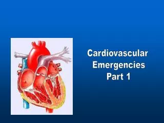
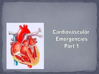
Cardiovascular Emergencies. Chapter 12. Cardiovascular Emergencies. Cardiovascular disease (CVD) claimed 931,108 lives in the US during 2001. 2,551 per day Almost two people per minute! CVD accounts for 38.5% of all deaths. One of every 2.6 deaths. Blood Flow Through the Heart.

Cardiovascular Emergencies
E N D
Presentation Transcript
Cardiovascular Emergencies Chapter 12
Cardiovascular Emergencies • Cardiovascular disease (CVD) claimed 931,108 lives in the US during 2001. • 2,551 per day • Almost two people per minute! • CVD accounts for 38.5% of all deaths. • One of every 2.6 deaths
Cardiac Compromise • Chest pain results from ischemia • Ischemic heart disease involves decreased blood flow to the heart. • If blood flow is not restored, the tissue dies.
Atherosclerosis • Materials build up inside blood vessels. • This decreases or obstructs blood flow. • Risk factors place a person at risk.
Angina Pectoris • Pain in chest that occurs when the heart does not receive enough oxygen • Typically crushing or squeezing pain • Rarely lasts longer than 15 minutes • Can be difficult to differentiate from heart attack
Heart Attack • Acute myocardial infarction (AMI) • Pain signals death of cells. • Opening the coronary artery within the first hour can prevent damage. • Immediate transport is essential.
Signs and Symptoms • Sudden onset of weakness, nausea, sweating without obvious cause • Chest pain/discomfort • Often crushing or squeezing • Does not change with each breath • Pain in lower jaw, arms, back, abdomen, or neck • Sudden arrhythmia with syncope • Shortness of breath or dyspnea • Pulmonary edema • Sudden death
Pain of Heart Attack • May or may not be caused by exertion • Does not resolve in a few minutes • Can last from 30 minutes to several hours • May not be relieved by rest or nitroglycerin
Sudden Death • 40% of AMI patients do not reach the hospital. • Heart may be twitching.
Arrhythmias • Bradycardia • Ventricular Tachycardia
Cardiogenic Shock • Heart lacks power to force blood through the circulatory system. • Onset may be immediate or not apparent for 24 hours after AMI.
Congestive Heart Failure • CHF occurs when ventricles are damaged. • Heart tries to compensate. • Increased heart rate • Enlarged left ventricle • Fluid backs up into lungs or body as heart fails to pump.
You are the provider • You are a volunteer EMT-B in a rural area. You are dispatched to an older man complaining of severe chest pain. • ALS has been dispatched. • You arrive to find the patient clutching his chest. The pain is the worst he has ever had. • The patient has nitroglycerin but has not taken it yet. • What is wrong with this patient? • What must you know before administering any medication? • What must you specifically know before assisting a patient with nitroglycerin?
Scene Size Up • Scene size-up • General impression • Is the patient responsive?
Initial assessment • Chief complaint on responsive patients • A chief complaint of chest discomfort, shortness of breath, or dizziness must be taken seriously. • Airway and breathing • Circulation
Transport Decision • Is the patient a life threat? • Stable patients • Transport in gentle manner. • Avoid lights and siren. • Do not let patient exert or strain self. • Specialty facilities
You are the provider • You obtain a brief history while taking the patient’s blood pressure. • Your partner retrieves the nitroglycerin and obtains permission from medical control. • Your partner administers the nitroglycerin. • What else can you do at this time?
Focused History and Physical Exam • SAMPLE • OPQRST • Medications are important! • Medications often prescribed for CHF: • Furosemide • Digoxin • Amiodarone
Focused Physical Exam • Cardiac and respiratory systems • Look for skin changes. • Lung sounds • Baseline vital signs • BOTH systolic and diastolic BP readings
Communication • Relay history, vital signs, changes, medications, and treatments.
Aspirin • Administer according to local protocol. • Prevents clots from becoming bigger • Normal dosage is from 162 to 324 mg.
Nitroglycerin • Forms • Pill, spray, skin patch • Effects • Relaxes blood vessel walls • Dilates coronary arteries • Reduces workload of heart
Nitroglycerin Contraindications • Systolic blood pressure of less than 100 mmHg • Head injury • Maximum dose taken in past hour • Use of ED medications
Nitroglycerin Potency • Nitroglycerin loses potency over time. • Especially if exposed to light • When nitroglycerin tablets lose potency: • May not feel the fizzing sensation • May not experience the burning sensation and headache • Fizzing only occurs with a potent tablet, not in the spray form
Assisting With Nitroglycerin • Obtain order from medical direction. • Take patient’s blood pressure. • Check that you have right medication, patient, and delivery route. • Check expiration date. • Find out last dose taken and effects. • Be prepared to lay the patient down. • Administer tablet or spray under tongue. • Have patient keep mouth closed until tablet dissolves or is absorbed. • Recheck blood pressure. • Record each activity and time of application. • Reevaluate and note response. • May repeat dose in 3 to 5 minutes.
Detailed Physical Exam • Perform if time allows. • Do not gather information unless: • Patient’s condition is stable • Everything else is done
Ongoing Assessment • Repeat initial assessment. • Reassess vital signs every 5 minutes. • Monitor closely. • If cardiac arrest occurs, begin defibrillation or CPR immediately. • Record interventions, instructions from medical control, patient’s response. • Obtain medical control physician’s signature.
You are the provider • ALS arrives and you report your interventions and vital signs. • ALS performs cardiac monitoring and prepares for morphine administration. • The patient’s pain is gone by the time you reach the hospital.
Heart Surgeries and Pacemakers • Coronary artery bypass graft (CABG) • Angioplasty • Cardiac pacemaker
Automatic Implantable Cardiac Defibrillators • Maintains a regular heart rhythm and rate • Do not place AED patches over pacemaker. • Monitor heart rhythm and deliver shocks as needed. • Low electricity will not affect rescuers.
Cardiac Arrest • The complete cessation of cardiac activity, either electrical, mechanical, or both.
Automated External Defibrillator (AED) • AEDs come in various models. • Some operator interaction required. • A specialized computer recognizes heart rhythms that require defibrillation.
Potential AED Problems • Battery is dead. • Patient is moving. • Patient is responsive and has a rapid pulse.
AED Advantages • ALS providers do not need to be on scene. • Remote, adhesive defibrillator pads are used. • Efficient transmission of electricity
Non-Shockable Rhythms • Asystole • Pulseless electrical activity
Rationale for Early Defibrillation • Early defibrillation is the third link in the chain of survival. • A patient in ventricular fibrillation needs to be defibrillated within 2 minutes.
AED Maintenance • Read operator’s manual. • Check AED and battery at beginning of each shift. • Get a checklist from the manufacturer. • Report any failures to the manufacturer and the FDA.
Medical Direction • Should approve protocols • Should review AED usage • Should review speed of defibrillation • Should provide review of skills every 3 to 6 months
Preparation • Make sure the electricity injures no one. • Do not defibrillate a patient lying in pooled water. • Dry a soaking wet patient’s chest first. • Do not defibrillate a patient who is touching metal. • Remove nitroglycerin patches. • Shave a hairy patient’s chest if needed.
Using an AED (1) • Assess responsiveness. • Stop CPR if in progress. • Check breathing and pulse. • If patient is unresponsive and not breathing adequately, give two slow ventilations. • If there is a delay in obtaining an AED, have your partner start or resume CPR. • If an AED is close at hand, prepare the AED pads. • Turn on the machine. • Remove clothing from the patient’s chest area. Apply pads to the chest. • Stop CPR. • State aloud, “Clear the patient.”
Using an AED (2) • Push the analyze button, if there is one. • Wait for the computer. • If shock is not needed, start CPR. • If shock is advised, make sure that no one is touching the patient. • Push the shock button. • After the shock is delivered, begin 5 cycles of CPR, beginning with chest compressions. • After 5 cycles, reanalyze patient’s rhythm. • If the machine advises a shock, clear the patient and push shock button. • If no shock advised, check for pulse. • If the patient has a pulse, check breathing. • If the patient is breathing adequately, provide oxygen via nonrebreathing mask and transport.
Using an AED (3) • If the patient is not breathing adequately, use necessary airway adjuncts and proper positioning to open airway. • Provide artificial ventilations with high-concentration oxygen. • Transport. • If the patient has no pulse, perform 2 minutes of CPR. • Gather additional information on the arrest event. • After 2 minutes of CPR, make sure no one is touching the patient. • Push the analyze button again (as applicable). • If necessary, repeat alternating CPR/Analyze/Shock until ALS arrives. • Transport and check with medical control. • Continue to support the patient as needed.
After AED Shocks • Check pulse. • No pulse, no shock advised • No pulse, shock advised • If a patient is breathing independently: • Administer oxygen. • Check pulse. • If a patient has a pulse but breathing is inadequate, assist ventilations.
Transport Considerations • Transport: • When patient regains pulse • After delivering six to nine shocks • After receiving three consecutive “no shock advised” messages • Keep AED attached. • Check pulse frequently. • Stop ambulance to use an AED.
Cardiac Arrest During Transport • Check unconscious patient’s pulse every 30 seconds. • If pulse is not present: • Stop the vehicle. • Perform CPR until AED is available. • Analyze rhythm. • Deliver shock(s). • Continue resuscitation according to local protocol. • If patient becomes unconscious during transport: • Check pulse. • Stop the vehicle. • Perform CPR until AED is available. • Analyze rhythm. • Deliver up to three shocks. • Continue resuscitation according to local protocol.