
Development of Cysteine-Linked Sorbitol Biosensor Using Gold Electrode
A novel biosensor based on sorbitol dehydrogenase linked to gold by cysteine was developed for early diagnosis of diabetic complications. The bioelectronic interface efficiently transfers electrons for sorbitol detection.

Development of Cysteine-Linked Sorbitol Biosensor Using Gold Electrode
E N D
Presentation Transcript
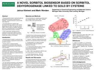
Sorbitol Fructose 2e- NAD+ Each of cysteine’s three different functional groups (shaded) form chemical bonds. 2e- Fig. 1: Molecular Structure of Cysteine ToluidineBlue O 3-Carboxy-phenylBoronic Acid 2e- Cysteine Gold Electrode A NOVEL SORBITOL BIOSENSOR BASED ON SORBITOL DEHYDROGENASE LINKED TO GOLD BY CYSTEINE Department of Chemical Engineering and Materials ScienceMichigan State University, East Lansing, Michigan Jairus Kleinert and Mark Worden • Abstract • Elevated sorbitol levels in red blood cells are associated with severe complications of diabetes. A sorbitol biosensor could provide early diagnosis. • A novel bioelectronic interface was developed that uses cysteine to link sorbitol dehydrogenase (SDH), its required cofactor (NAD+), and an electron mediator (toluidine blue O) to a gold electrode in an arrangement that provides efficient electron transfer. • Cyclic voltammetry indicated that the interface functions as an effective sorbitol biosensor, in which the peak current varies linearly with sorbitol concentration. • Introduction • Elevated levels of sorbitol in red blood cells can signal diabetic complications before other symptoms appear.1 • A sensor able to measure sorbitol concentrations could allow early diagnosis, treatment, and prevention of vision loss and nerve damage.2 • Sorbitol dehydrogenase enzymatically oxidizes sorbitol to fructose. Electrons produced in the reaction could be transferred to an electrode, resulting in a electrochemical biosensor for sorbitol. • Sorbitol dehydrogenase transfers electrons from sorbitol to an electron-carrying cofactor (NAD+). Achieving efficient electron transfer from NAD+ to the electrode requires an electron mediator. • Our lab recently developed a novel approach that uses the amino acid cysteine (Fig. 1) to electrically couple NAD+ and an electron mediator to a gold electrode.3 The goal of this study is to apply this approach to develop a sorbitol biosensor. • Materials and Methods • A novel bioelectronic interface was fabricated that includes cysteine, SDH, NAD+, toluidine blue O (TBO), and a gold electrode. • Cysteine’s three functional groups allow the molecular components of the interface to be bound in a unique conformation (Fig. 2). • 1. Cysteine: Cysteine’s sulfhydryl group was attached to the gold electrode by a thiol bond. • 2. Toluidine Blue O: An ester bond was formed between TBO and cysteine’s carboxylic acid group. • 3. 3-Carboxyphenyl Boronic Acid: An ester bond was formed between CBA and cysteine’s amine group. • 4. NAD+: NAD+ was attached to CBA through the cis-diol group on both molecules. • Sorbitol Dehydrogenase: SDH was attached to NAD+ by an affinity linkage. • Results and Discussion • The electrochemical activity of the interface was studied using cyclic voltammetry (CV) in aqueous sorbitol solutions. Each curve in Fig. 3 is for a different sorbitol concentration. • Peak CV current varied linearly with sorbitol concentration, indicating that the interface serves as an effective sorbitol biosensor. Conclusions Cysteine was used to attach SDH, NAD+, and TBO to a gold electrode in a unique conformation. The resulting bioelectronic interface functions as a sorbitol biosensor. References 1. Ciulla, T.A.; Amador, A. G.; Zinman B. “Diabetic retinopathy and diabetic macular edema -- Pathophysiology, screening, and novel therapies.” Diabetes Care. 26 (2003) 2653-2664. 2. Kwang-Hyok, S., et. al. “A sensitive assay of red blood cell sorbitol level by high performance liquid chromatography: potential for diagnostic evaluation of diabetes.” Clinica Chimica Acta. 354 (2005) 41-47. 3. Hassler, B.; Worden, R. “Versatile bioelectronic interfaces based on heterotrifunctional linking molecules.” Biosensors and Bioelectronics, in Press. Acknowledgements This work was supported by the Michigan Technology Tri-Corridor Grant to the Center for Nanostructured Biomimetic Interfaces (www.biomimetic.org). The Kikkoman Corporation donated the SDH enzyme. Fig. 4: Calibration Curve for Sorbitol Biosensor Fig. 2: Schematic Diagram of Biosensor Fig. 3: CV Curves for Sorbitol Biosensor


