BRAIN ATTACK
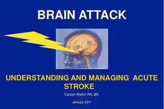
BRAIN ATTACK. UNDERSTANDING AND MANAGING ACUTE STROKE. Carolyn Walker RN, BN. January 2011. Brain Attack: Understanding and Managing Acute Stroke. Learning Objectives : Upon completion of this session, participants will be able to: Describe the 2 major types of stroke

BRAIN ATTACK
E N D
Presentation Transcript
BRAIN ATTACK UNDERSTANDING AND MANAGING ACUTE STROKE Carolyn Walker RN, BN. January 2011
Brain Attack: Understanding and Managing Acute Stroke Learning Objectives: Upon completion of this session, participants will be able to: • Describe the 2 major types of stroke • Identify the location of stroke given stroke symptoms • Describe the management of hypertension in acute stroke • Explain the appropriate management of acute ischemic stroke
Epidemiology of Stroke: The Canadian Perspective • 50,000 new stroke patients/year in Canada† • 5,500 Albertans suffer a stroke each year • Every 10 minutes someone in Canada suffers a “brain attack” • 3rd leading cause of death in Canada • The leading cause of adult disability • 200,000–300,000 stroke survivors† • Cost to society: $300-400 million/yr Alberta • 28% of stroke patients are under age 65* †Statistics Canada
What is a stroke? BLOCKAGEBREAKAGE blood vessel occlusion or blood vessel rupture (clot / atherosclerosis) sudden interruption in cerebral blood flow brain injury to affected area brain death of affected area
Stroke: Brain Attack • Stroke is a “brain attack” • Stroke is an EMERGENCY!
Frequency of Stroke by Type • Ischemic (85%) Thrombotic (54%), Embolic (31%) • Ischemic Stroke – 65% • TIA – 20% • symptoms resolve • no brain cell death • 20-40% of strokes are proceeded by TIA • Hemorrhagic (15%) • Intracerebral – 10% • Subarachnoid – 5% Blockage Breakage
The Brain • Cerebrum • Diencephalon • Cerebellum • Brainstem
Cerebrum • Center for highest function • Governs thought, memory, reasoning, sensation and voluntary movement • Divided into two hemispheres • Left Hemisphere • dominant in 95% of people • Right Hemisphere
Functions of Cerebral Hemispheres PHOTO: Courtesy of National Stroke Association
Motor and Sensory Function PHOTO: Courtesy of National Stroke Association
Cerebrum • Basal ganglia • Bands of grey matter deep within the cerebral hemispheres • Control automatic associated movements • i.e. arm swing alternating with leg movement • posture
Diencephalon • Includes thalamus and hypothalamus • Extends from cerebrum to midbrain • Surrounds 3rd ventricle • Thalamus • Receives sensory input • Relay station to cerebral cortex • Hypothalamus • Major control centre • Regulation of temp, H2O balance, sleep, behavior • Coordinator of autonomic nervous system activity
Cerebellum • Located under occipital lobe • Unconscious motor coordination of voluntary movement • i.e. complex coordination of different muscles needed to juggle, swim, etc. • Equilibrium • Muscle tone
Brain Stem • Central core of brain • Consists mostly of nerve fibers • Midbrain • Auditory/visual systems • Pons • Respiratory centers • Medulla • Respiratory and vasomotor control
Blood Supply to the Brain PHOTO: Courtesy of National Stroke Association
Blood Supply to the Brain 90% of all strokes • Carotid Arteries & Branches: anterior 2/3 cerebral of hemispheres • Vertebral Arteries & Branches: posterior and medial regions of hemispheres brainstem diencephalon (thalamus/hypothalamus) cerebellum Courtesy Genentech 10% of all strokes
Hemorrhagic Stroke Intracerebral Hemorrhage Subarachnoid hemorrhage
Intracerebral Hemorrhage • Result of ruptured Blood vessel • Hypertension most common cause • Usual Presentation: • Headache • Hemiplegia • Decreased level of consciousness • Nausea & Vomiting
Subarachnoid Hemorrhage • Blood vessel ruptures & bleeds into subarachnoid space (Aneurysms/arteriovenous malformations ) • “Worst headache of one’s life” • Nausea & vomiting • Neck stiffness • Neurologic signs don’t fit pattern of a single blood vessel • Varying level of consciousness
Management of SAH and ICH:The First Few Hours • Correct airway, breathing or circulation • Treat severe elevation of BP • Obtain neurosurgical consult • Treat elevated intracranial pressure • Admin anticonvulsant therapy if seizures
Intracerebral Hemorrhage: Hypertension Management Recommendations: Maintain SBP < 180 mmHg and DBP < 100 mmHg • MAP < 130 mmHg if history of hypertension DO NOT REDUCE BP BY MORE THAN 20% CONTACT STROKE SPECIALIST AT COMPREHENSIVE STROKE CENTER!
Etiology of Ischemic Stroke Graphics courtesy Boehringer Ingelheim
Classifications of Ischemic Stroke • Small vessel disease • Lacunar infarction • Large vessel disease • Artery to artery emboli (large artery atherosclerosis) • Cardioembolic • Cryptogenic (Don’t know the Cause) • Other (Cocaine, coagulopathies)
Progression of Ischemic Stroke Graphics courtesy Boehringer Ingelheim
TIME IS BRAIN!In a typical large vessel acute ischemic stroke…- 1.9 million neurons - 14 billion synapses - 12 km of myelinatedfibersare destroyed each minute …(JL Saver, 2006)
Symptoms of “Brain Attack” Speech Strength Sight
ACUTE STROKE OUTCOMES CAN BE IMPROVED IF WE PROVIDE ARAPID COORDINATED RESPONSE!
Approaches to Acute Therapy • Neuroprotection • Studies* • Reperfusion
REPERFUSION - Thrombolytic Agents • Intravenous rt-PA • Strict protocols for use with ischemic stroke • Improves outcomes compared to the risk of serious bleeding
Canadian Stroke Strategy:Best Practice Recommendations 2010 • All patients with disabling acute ischemic stroke who can be treated within 4.5 hours after symptom onset should be evaluated without delay to determine their eligibility for treatment with t-PA.
Diminishing Returns over Time Favorable Outcome (mRS 0-1, BI 95-100, NIHH 0-1) at Day 90 Adjusted odds ratio with 95% confidence interval by stroke onset to treatment time (OTT) ITT population (N=2776) Pooled Analysis NINDS tPA, ATLANTIS, ECASS-I, ECASS-II Courtesy Brott T et al
REPERFUSION • Intra-arterial lytic • ultrasonic clot-busting
REPERFUSION: Devices - Clot Retrieval Mechanical Thrombectomy Devices • MERCI study: MERCI device Mechanical Embolus Removal in Cerebral Ischemia • Penumbra device
Canadian Stroke Strategy:Best Practice Recommendations 2010 There remain situations where there are sparse or little clinical trial data to support the use of thrombolytic therapy: • Paediatric stroke • Over 80 years with diabetes • Present within time window but do not meet current criteria for treatment with IV t-PA • Intra-arterial thrombolysis Treat based on clinical decision of physician and family
EMS Protocol- Arrival at scene PRIORITY IS LOAD AND GO ABC’s first Determine time last known to be normal Acute Stroke Screen Perform directed neurological assessment Blockage or Breakage?
Onset Time • Onset Time = Time when patient was last seen well • Requires detective skills
Pre-Hospital Care:Direct transport to Primary Stroke Centre (PSC) • A standardized acute stroke diagnostic screening tool should be used by paramedics • Pts with symptoms of stroke should be transported without delay to the closest institution that provides emergency stroke care • Direct transport protocols must be in place • Paramedics must notify the receiving facility • Transfer care to receiving facility without delay (scene time < 10 min) EMS Stroke Screening Form
0 10 20 30 40 50 60 70 80 90 minutes
vs CT scanner 40 miles 8 miles Local hospital No CT scanner intraclot lysis ICH evacuation vs 70 miles Early ICA revascularization 170 miles Interventional Facilities- interventional neurorad, neurosurgery Comprehensive Stroke Center vs Helical or multislice CT scanner 24h/365d coverage Primary Stroke Center
Alberta Stroke Centre Locations • Primary Stroke Centre (PSC): 14 • CT scan availability • Door to CT < 20 min. with a pre-alert • Stroke expertise on-site or available by Telestroke link • r-tPA treatment availability • May not be available 24/7 • Comprehensive Stroke Centre (CSC): 3 • CT scan availability • Door to CT < 20 minutes with a pre-alert • Stroke team on-site • Neurological expertise on-site • Neurointerventional expertise on-site • Central hub of stroke Neurologist expertise in a telestroke network
Initial Management of Stroke:A. Immediate General Assessment • Assess A B C’s, vital signs (BP, HR, Temp***) • Provide oxygen (O2 sats >95%, if COPD >90%) • Start an IV Line (large bore)- no dextrose • 12 Lead ECG / cardiac monitoring • Obtain blood samples (CBC, lytes, Cr, gluc, PTT, INR) • Check Blood Sugar Levels*** • Perform general neurological screening • Alert Stroke Team
Canadian Stroke Strategy:Best Practice Recommendations 2010 • Monitoring in the acute phase should include • HR and rhythm, BP, temp, O2 sat, hydration, swallowing ability and presence of seizure activity • Initial blood work should include • CBC, lytes, Cr, urea, glucose, INR, PTT, TSH, fasting lipids, CK and troponin • Neurovascular Imaging – should undergo brain imaging (MRI or CT) immediately • Vascular imaging of the brain and neck arteries ASAP • Cardiovascular investigations • After initial ECG-daily ECG’s x 72 hrs • May also monitor x 72 hrs to detect afib • Echocardiography if suspect embolic stroke
Canadian Stroke Strategy:Best Practice Recommendations 2010 • Acute Aspirin Therapy • All stroke pts not on antiplatelet therapy should be given at least 160 mg of ASA immediately as a one time loading dose after brain imaging excludes hemorrhage • If treated with t_PA- delay ASA until after 24 hour CT excluding hemorrhage • If taking ASA may consider plavix
Hypertension During Acute Stroke • Systolic BP > 160mmHg is seen in over 60% stroke patients (Robinson et al, Cerebrovasc Dis., 1997) • Often transient, lasting 24-72 hours and in most patients does not require treatment. • Little evidence and no benefit seen for rapid lowering of BP in acute stroke without rt-PA