Download
1 / 13
130 likes | 317 Views
Cytology & The Discovery of Cells. Packet #14 Chapter #6. Cell Theory. Cell Theory. Scheleiden and Schwann stated that cells are the fundamental unit of life. Virchow stated that cells arise from preexisting cells. Weismann described that cells have a “common ancestor.”

E N D
Cytology & The Discovery of Cells Packet #14 Chapter #6
Cell Theory Scheleiden and Schwann stated that cells are the fundamental unit of life. Virchow stated that cells arise from preexisting cells. Weismann described that cells have a “common ancestor.” These three thoughts compose the cell theory.
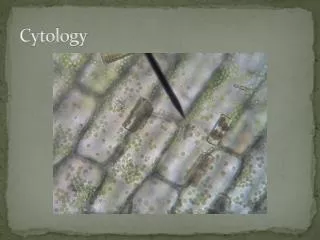
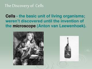
Cytology Began with Robert Hooke and the light microscope in the early 1900’s. Hooke is credited with seeing the first cells; rather, he first identified the cell walls of dead cells
Light Microscopes • Light microscopes, more commonly used to study stained or living cells, first identified organelles in the early 1900’s • Discovery made using different stains. • Phase and contrast microscopes allowed unstained living cells to be observed. • Fluorescence microscopes can identify the location of molecules within cells. • Light microscopes, though useful, are limited by their resolution power.
Electron Microscopes • Electron microscopes became available allowing cells, and their contents, to be greatly magnified. • TEM {Transmission Electron Microscope} • Visualization of structures within sections of tissues • SEM {Scanning Electron Microscope} • Visualization of entire specimens
Cell Fractionation • Cell fractionation allowed the study of cell components. • Involves centrifugation • Differential centrifugation separate cellular components based on size and density. • Density gradient centrifugation allows further purification.
Introduction The discovery of cells, and their components, led to the distinction between prokaryotic cells and eukaryotic cells.
Prokaryotic Cells Prokaryotes are structurally simpler than eukaryotic cells. Prokaryotic cells lack membrane-bound organelles and are typically smaller than eukaryotic cells. Prokaryotic cells have a plasma membrane and typically a cell wall. In prokaryotes, DNA is located in the nuclear area or nucleoid. Prokaryotes have ribosomes and storage granules.
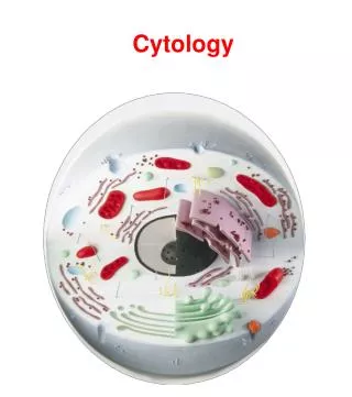
Eukaryotic Cells • Characterized by membrane bound organelles. • Increasing cell size allows increased specialization in eukaryotes. • Some organelles are only found in certain eukaryotic cells • Others are common to most or all eukaryotic cells.
Prokaryotes vs. Eukaryotes Prokaryotic Cells Eukaryotic Cells • No distinct nucleus • No chromosomes • Circular strands of DNA known as plastids • No membrane bound organelles • Ribosomes are smaller • Flagella, if present, lack internal 9 + 2 fibril arrangement • No mitosis or meiosis occurs Distinct, membrane-bounded nucleus Chromosomes present on which DNA is located Chloroplasts and mitochondria may be present Ribosomes are larger Flagella have 9 + 2 internal fibril arrangement Mitosis and/or meiosis occurs