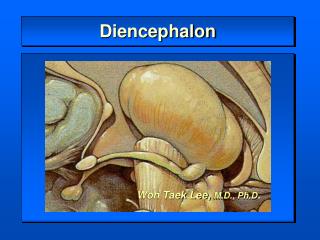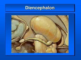
DIENCEPHALON-HYPOTHALAMUS
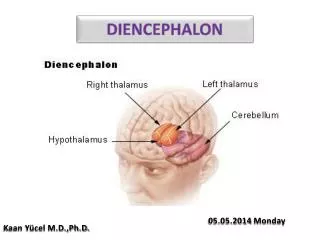
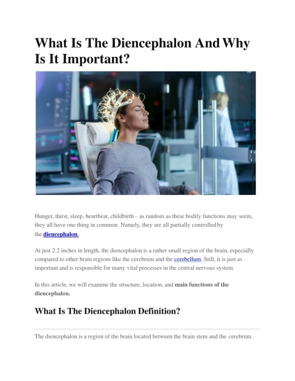
DIENCEPHALON-HYPOTHALAMUS. Diencephalon [“ between brain”]. Mostly hidden from view Between cerebral hemispheres 2% of CNS by weight Widespread and important sensory connections. Majority of sensory, motor and limbic pathways involve one or more stops in this region


DIENCEPHALON-HYPOTHALAMUS
E N D
Presentation Transcript
Diencephalon [“ between brain”] • Mostly hidden from view • Between cerebral hemispheres • 2% of CNS by weight • Widespread and important sensory connections
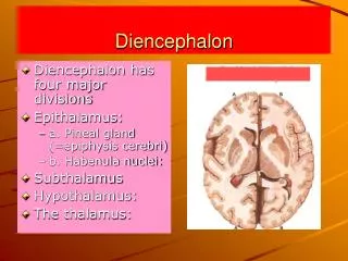
Majority of sensory, motor and limbic pathways involve one or more stops in this region • 4 parts – each part includes the term ‘thalamus’ [ inner chamber]
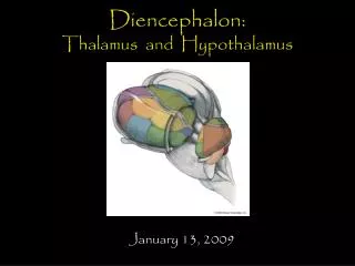
Epithalamus –including pineal gland and few nearby neural structures • Dorsal thalamus=thalamus • Subthalamus • Hypothalamus
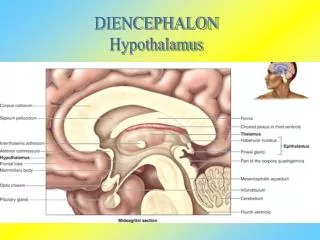
Visible part of diencephalon is inferior surface of hypothalamus • Includes mammillary bodies and infundibulum • Entire medial surface is wall of 3rd ventricle, visible in a hemisected brain
Superiorly, it borders body of lateral ventricle • Laterally- internal capsule • Caudal boundary-plane through posterior commissure and caudal edge of mammillary bodies • Rostral boundary-plane through back of anterior commissure and front of optic chiasm
Boundaries are approximate • Neural tissue is continuous across boundaries • Certain thalamic nuclei protrude through posterior boundary to a position alongside midbrain
Epithalamus • Includes pineal gland and habenular nuclei
Pineal gland • Midline, unpaired • Resembles a pine cone • Rostral to superior colliculi • Once considered to be the seat of the soul
Pineal tumours compress midbrain leading to • Hydrocephalus • Deficits in eye movements and pupillary reactions • Altered sexual development
Receives light – regulated input by a circuitous pathway • Retina →hypothalamus→ intermediolateral cell column→ postganglionic fibres of superior cervical ganglion→pineal gland • No known neural output
Secretes a hormone- melatonin [derived from serotonin] • Secretion increases during darkness • Related in humans to sleep-wake cycles • Gland undergoes calcification after the age of 17
Calcified gland is a useful radiologic landmark • Slight shifts in pineal position can be indicative of expanding masses of different types
Basic facts • Small part of diencephalon [ 4g in weight] • Important as a nodal point in pathways concerned with autonomic, endocrine, emotional and somatic functions designed to promote homeostasis
Widespread sets of connections • Various components of limbic system • Outputs influencing pituitary gland • Interconnections with various visceral and somatic nuclei[ motor and sensory,of brainstem and spinal cord]
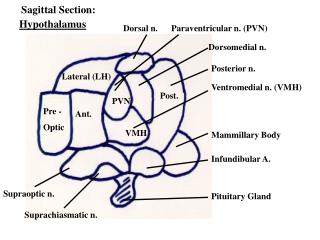
Boundaries of inferior surface • Optic tracts, optic chiasma, mammillary bodies • This area exclusive of mammillary bodies is called tuber cinerium [‘gray swelling’] • Medial eminence protrudes from surface of tuber cinerium , and is continuous with infundibular stalk, which in turn is continuous with posterior lobe of pituitary
Infundibular stalk +posterior lobe of pituitary=neurohypophysis
Medial surface • Anterior extent-lamina terminalis • Superiorly- hypothalamic sulcus • Posteriorly- caudal edge of diencephalon
Neural tissue anterior to a plane passing through anterior edge of optic chiasma and posterior edge of anterior commissure is functionally continuous with hypothalamus=preoptic area • Considered a part of anterior hypothalamus
Divisions [anteroposterior] • Anterior • Tuberal • Posterior
Anterior region- above optic chiasma • Tuberal – above and including tuber cinerium • Posterior – above and including mammillary bodies
Parasagittalsection.Regions ;a-anterior, p-posterior,t-tuberal, po-preoptic
Longitudinal zones • Periventricular- in the wall of 3rd ventricle [rostral continuation of PAG] • Lateral –lateral to fornix • Medial zone [in between the two] –populated by series of hypothalamic nuclei • The 1st 2 zones contain neurons and are avenues via which ascending and descending axons enter, leave or traverse hypothalamus
Coronal section.medial-lateral subdivisions. L-lateral, m-medial,pe-periventricular
Periventricular zone • Traversed by dorsal longitudinal fasciculus[bundle of hypothalamic afferents and efferents] • Contains suprachiasmatic and arcuate nuclei • Suprachiasmatic – tiny – less than 1 mm square and fewer than 10,000 neurons • ‘master clock’ for our circadian rhythms
Receives direct retinal projections which entrain it to the actual day length • Its neurons also contain melatonin receptors • Night-time rise in pineal melatonin secretion probably helps ‘set’ the circadian rhythm
Lateral zone • Mainly scattered cells interspersed among longitudinally running fibers of Medial forebrain bundle • Anteriorly- continuous with lateral preoptic nucleus- an important sleep-promoting area • Caudally- continuous with midbrain reticular formation
Also has • Parts of supraoptic nucleus • Lateral tuberal nuclei • Tuberomammillary nucleus [source of histaminergic fibers that project widely to cerebral cortexand thalamus-participate in sleep-wake cycles]