Download
1 / 42
420 likes | 564 Views
KLINIKA ANESTÉZIOLÓGIE A INTENZÍVNEJ MEDICÍNY LF UPJŠ A FNLP KOŠICE. C oma, head injury. Judita Capkova, MD. PhD. Jozef Firment, MD. PhD. Department of Anaesthesiology & Intensive Care Medicine Šafárik University Faculty of Medicine, Košice. Coma.

E N D
KLINIKA ANESTÉZIOLÓGIE A INTENZÍVNEJ MEDICÍNY LF UPJŠ A FNLP KOŠICE Coma,head injury Judita Capkova, MD. PhD. Jozef Firment, MD. PhD. Department ofAnaesthesiology & Intensive Care MedicineŠafárik University Faculty of Medicine, Košice
Coma Is a „state of unarousable unresposiviness“(of unconsciousness from which the patient cannot be aroused) • No evidence of arousal: no spontaneous eye opening, no speech, voluntary limb movement • Unresponsive to external stimuli, although abnormal postures may be • Involuntary movements (seizures) may occur • GCS – level of consciousness, coma: GCS ≤ 8
GLASGOW COMA SCALE Decorticate posturing Decerebrate posturing
Metabolic Toxic Infection with or Structural lesions without Focal brainstem signs Lateralizing cerebral signs Meningeal irritation Causes of coma • toxic, metabolic causes usually do not produce focal signs- infections, structural lesions producefocal signs
Coma without focal/lateralizing neurological signs • Anoxia/ hypoperfusion • Metabolic: e.g. Hypo/-hyperglycaemia, acidosis/alkalosis, hepatic or renal failure • Intoxications: e.g. alcohol, opiates,benzodiazepines,.. • Endocrine : hypothyreoidism • Hypo- or hyperthermia • Epilepsy • Hypertensive encephalopathy
Coma with focal/lateralizing neurological signs ( due to brainstem or cerebral dysfunction) • Vascular : cerebral haemorrhage or infarction • Supra or infratentorial space-occupying lesion: tumour, haematoma, abscessComa with meningism • Meningitis, encephalitis • Subarachnoid haemorrhage
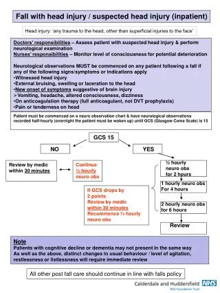
Immediate management 1.Stabilize the patient ABC • Open the airway, breathing.give oxygen, stabilise the cervical spine as required • OTI, ventilation ? (GCS ≤ 8) pO2, pCO2 • Support the circulation: correct hypotension (colloids, inotropes), CVP? • Treat seizures (diazepam, phenytoin) - • Take blood for glucose, U+Es, calcium, liver enzymes, albumin, clotting screen,FBC, toxicology (+urine) CMRO2
2. Consider giving:thiamine(Wernickes encephalopathy) glucose (40ml 40% glucose) naloxon(opiate intoxication)flumazenil(benzodiazepine intoxication) Hypoglycaemia
3. Examine patient: History General examination • Core temperature,heart rate, rhythm, BP, respiratory pattern, breath, skin, heart, abdomen, fundi Is there meningism? – neck stiffness (inflammation, blood) Asses GCS Look for evidence of brainstem dysfunction Are there lateralizing signs?
Test brainstem dysfunction • Pupillary response • Corneal reflex • Spontaneous eye movements • Oculocephalic response/Doll’s headmanoeuvre • Oculovestibular response • Swallowing reflex • Respiratory pattern
Motor function: • Decorticate posturing – lesions above the pons • Decerebrate posturing – pontine damage Decorticate posturing Decerebrate posturing
4. Plan for further investigations: 1. Brainstem function intact:urgent CT head scan : - lesions (subdural haematoma,..),- normal – lumbar puncture, CSF analysis
4. Plan for further investigations: 1. Brainstem function intact:urgent CT head scan : - lesions (subdural haematoma,..),- normal – lumbar puncture, CSF analysis 2.Brainstem function not intact:Signs ICH (intracranial hypertension): - early: headache,vomiting,seizures, focal neurology, papilloedema- late: incr. BP, bradycardia, coma, Cheyne Stokes breathing, apnoe. - ifherniation syndromeappears to be progressing rapidly - mannitol, hyperventilation, surgeon- if herniation syndrome appears to be progressing not so rapidly – mannitol and CT, surgeon
4. Plan for further investigations: 1. Brainstem function intact:urgent CT head scan : - lesions (subdural haematoma,..),- normal – lumbar puncture, CSF analysis 2.Brainstem function not intact: - ifherniation syndromeappears to be progressing rapidly - mannitol, hyperventilation, surgeon- if herniation syndrome appears to be progressing not so rapidly – mannitol and CT Hyperventilation- hypocapnia- vasoconstrictionofcerebralaa. – decreaseofintracranialpressure
Head injury (HI) • Primary brain injury: - brain lacerations, contusions, diffuse axonal injury due to accelaration or deceleration - the neurones lost at the time of HI are lost forever
Secondary injury: • Due to raised intracranial pressure (ICP) andinadequate cerebral perfusion • Causes of secondary brain injury : Intracranial: • Haematoma (extradural, subdural,intracerebral) • Brain swelling/ oedema • Cerebral ischemia (vasospasm, seizures) • Inflammatory mediators Systemic : • Hypoxaemia • Hypotension • Hypercarbia • Severe hypocapnia • Pyrexia,.. • Anaemia • Hyper/hypoglycaemia
Prevention of secondary injury is the aim of the treatment.Prevention therapy may improve outcome.
De-compensation phase PRESSURE [mmHg] Transition phase 40 20 0 Compensation Phase VOLUME INTRACRANIAL PRESSURE (ICP) Up to 15 mmHg, above 40 malignant oedema
INCREASED ICP • Normal ICP 0-10 mmHg • ICP > 15-20 mmHg treatment is required • Causes of raised ICP: • Increased extracellular fluid: cerebral oedema • Increased cerebral blood flow : hypoxia, hypercarbia,..(vasodilatation) • Increased cerebral venous volume : venous obstruction in the neck, coughing,.. • Increased CSF volume : hydrocephalus,...
Patients with head injuries usually have a mixed type of oedema: vasogenic and cytotoxic.
Increased ICP >25 mmHg • ICP peaks at 72 h • Cerebral herniation • Reduced CPP(cerebral perfusion pressure)MAP – ICP = CPP causing ischemiaTherapy aim: CPP > 60 mmHg
Cerebral herniation Supratentorial herniation 1. Uncal 2. Central (transtentorial) 3. Cingulate (subfalcine) 4. Transcalvarial Infratentorial herniation 5. Upward (upward cerebellar or upward transtentorial) 6. Tonsillar (downward cerebellar)
Normally CBF (cerebral blood flow) is maintained constant by autoregulation between a MAP 50- 140 mmHg(MAP = APd + 1/3 (APs-APd)mean AP = diastolic AP + 1/3 (systolic AP- diastolic AP)
Autoregulation is impaired : head injury, acidosis (hypoxia, hypercarbia) • CBF varies passively with CPP (ischemia!!)
Hypoxia and hypercapnia • Dilates normal vessels and divert CBF away from damaged cerebral tissue • CBV (cerebral blood volume) and ICP- CPP and CBF- aggravates ischaemiain damaged brain tissue
Hypocapnia • Constricts normal vessels CBV and ICP CPP and CBF • !! Severe hypocapnia – exccess vasoconstriction – ischaemia in normal tissueRecommended: normal Pa CO24,6 – 5,3 kPa (35-40mmHg)
Raised ICP: immediate management • Open the airway, intubation, mechanical ventilation, keep Pa CO2 3,3 – 4,0 kPa (25-30mmHg) • Correct hypotension: colloids, infusions of inotropesCPP <70 mmHg is critical ! • Spinal immobilisation- all pt • Detect other injuries: 50% have potentially lethal thoracic or abdominal injuries • Treat seizures(increase O2consumption) • Sedation (paralysis) prevent ICP elevation in agitated pt • Take blood for glucose, U+Es, calcium, liver enzymes, albumin, clotting screen,FBC
Radiographic evaluation: • Immediate CT scan- in coma, GCS ≤ 8- GCS 9-13 with skull fractures • Intracranial hematoma is 10 x more common after skull fractures
Monitoring • GCS isadequate in mildinjuries • ICP intracranialpressure– severe HI • CerebraloxygensaturationSjO2 jugularvenousbulbfibreopthiccatheterSjO2< 55% inadequate (low) CBF
Normal curve shape Low compliance INTRACRANIAL PRESSURE
Management • Prevention of secondary injury is the aim:optimise CBF: MAP – ICP = CPP MAP>70 mmHg ICP < 15 mmHgCPP > 60 mmHg and oxygenation:SatO2 > 90%, SjO2 >55%
1. Reduce ICP: • Hyperventilation :PaCO23,3 – 4 kPa (25-30mmHg) not routinelly only if herniation appears • Loop diuretics (furosemid 20-40mg i.v.), osmotic agents (mannitol 0,5-1 g/kg )- reduce ICP • Improved venous drainage: midline haed position + 30°elevation, !! suctioning, PEEP, physiotherapy increase thoracic venous p. • Ventriculostomy drainage/decompressive surgery – if other fails • No corticosteroids
2. Reduce cerebral metabolism: • Avoid hyperglycaemia (BS 4-7 mmol/l) hyperglycaemia increase cerebral lactate production • Prophylactic anticonvulsants • Adequate analgesia and sedation: benzodiazepines, propofol, thiopentone • Antipyretics and cooling (33-34 °C maybe neuroprotective)
Treatcomplications: • Hypotalamicinjury :inappropriate ADH secretion – diabetes insipidus • Meningitis – ATB • Avoidnasogastictubes in basilarskullfracture
TBI, maxillofaciálne poranenie, haemothoraxTracheostómia – UVP, PEG, drenáž hrudníka
Thank you! jcapkova@capko.sk
TRAUMATIC BRAIN INJURY Hypoxia and acidosis Cerebral oedema
Immediate management • Stabilize the patient: ABCgive oxygen, support circulation, treat seizures, stabilise the cervical spine as required • Consider giving thiamine, glucose (40ml 40% glucose), naloxon, flumazenil • Examine patient • Plan for further investigations
ICP peaks at 72 h • CPP(cerebral perfusion pressure) = MAP - ICP • MAP = APd + 1/3 (APs-APd) • CPP is the effective pressure that results in blood flow to the brain.
CPP(cerebral perfusion pressure) = MAP - ICP • CBF (cerebral blood flow) is maintained constant by autoregulation (between a MAP 50- 140 mmHg).Autoregulation is impaired : head injury, acidosis (hypoxia, hypercarbia)CBF varies passively with CPP (ischemia!!) Therapy aim: CPP <70 mmHg is critical !
5. Progress in monitoring • Regular and frequent observations of vital signs and neurological state • Emergency treatment of raised ICP (intracranial pressure) Signs ICH (intracranial hypertension): - early: headache,vomiting,seizures, focal neurology, papilloedema- late: incr. BP, bradycardia, coma, Cheyne Stokes breathing, apnoe.