Head Injury
Head Injury. Head Injury. A head injury is any trauma that leads to injury of the scalp, skull, or brain. The injuries can range from a minor bump on the skull to serious brain injury.

Head Injury
E N D
Presentation Transcript
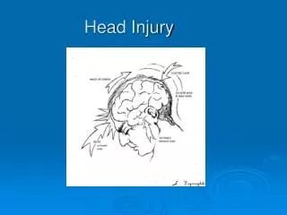
Head Injury • A head injury is any trauma that leads to injury of the scalp, skull, or brain. The injuries can range from a minor bump on the skull to serious brain injury. • Subarachnoid hemorrhage is bleeding in the area between the brain and the thin tissues that cover the brain. This area is called the subarachnoid space.
Hemorrhagic strokes • Hemorrhagic strokes include bleeding within the brain (intracerebral hemorrhage) and bleeding between the inner and outer layers of the tissue covering the brain (subarachnoid hemorrhage). • There are two main types of hemorrhagic strokes: intracerebral hemorrhage and subarachnoid hemorrhage. Other disorders that involve bleeding inside the skull include epidural and subdural hematomas, which are usually caused by a head injury.
Symptoms • Persistent headache, fluctuating drowsiness, confusion, memory changes, paralysis on the side of the body opposite the hematoma, and speech or language impairment. • In infants, a subdural hematoma can cause the head to enlarge (as in hydrocephalus), because the skull is soft and pliable. Therefore, pressure within the skull increases less in infants than it does in older children and adults.
Symptoms/Diagnosis • Chronic subdural hematomas are more difficult to diagnose because of the length of time between the injury and the development of symptoms. An older person with gradually developing symptoms, such as memory impairment and drowsiness, may be mistakenly thought to have dementia. CT can detect acute, subacute, and many chronic subdural hematomas. Magnetic resonance imaging (MRI) is particularly accurate for diagnosis of chronic subdural hematomas.
Treatments • Small subdural hematomas in adults do not require treatment because the blood is absorbed on its own. • Large hematomas causing symptoms such as persisting headache, fluctuating drowsiness, confusion, memory changes, and paralysis on the opposite side of the body, are drained surgically by drilling a small hole in the skull (Bur Hole). A drain is usually inserted and left in place for several days. The person is monitored closely for recurrences. In infants, doctors usually drain the hematoma for cosmetic if for no other reasons. • Only about 50% of people who are treated for a large acute subdural hematoma survive. People who are treated for a chronic subdural hematoma usually improve or do not worsen.
Intracerebral Hemorrhage • An intracerebral hemorrhage is bleeding within the brain. • Intracerebral hemorrhage usually results from chronic high blood pressure. • The first symptom is often a severe headache. • Diagnosis is based on symptoms and results of a physical examination and imaging tests. • Treatment may include vitamin K, transfusions, and, rarely, surgery to remove the accumulated blood.
Hyperventilation/ICP • As part of intensive treatment of traumatic brain injury, intracranial pressure (ICP) should be controlled when the cerebral perfusion pressure (CPP) falls below 70mmHg and/or the ICP is greater than 20mmHg. • Intracranial hypertension occurs in approximately 40% of all patients with severe traumatic brain injury. • There are several methods for controlling ICP. These are usually applied in a stepwise fashion to achieve control, where possible. The absolute requirement for the potentially severely brain injured patient is intubation. This protects and maintains the airway and allows for maximal oxygenation and control of ventilation. • Normal ICP values in adults are < 10 mm Hg, and a threshold of 20 mm Hg is usually accepted for starting active treatment.
Hyperventilation/ICP • The use of hyperventilation in the treatment of patients with TBI remains controversial. • Carbon dioxide dilates the cerebral blood vessels, increasing the volume of blood in the intracranial vault and therefore increasing ICP. • Mannitol should be reserved for acute control of ICP and administered in bolus form.
Hyperventilation/ICP • Previously, hyperventilation was used routinely to maximally reduce PaCO2. No studies have shown this to improve outcome in these patients. • Additionally, transcranial doppler (TCD) assessment and positron emission tomography (PET) shows this can induce significant constriction of cerebral vessels and this increase in cerebral vascular resistance may reduce cerebral blood flow to below the ischaemic threshold.
Fluids • Patients with severe brain injury should be kept normovolaemic. Previous regimens recommending that patients be kept 'dry' have essentially been discarded as there is significant risk of both hypotensive episodes (leading to a fall in cerebral perfusion) and systemic inflammatory response syndrome (SIRS) or multiple organ failure (MOF) leading to failure of oxygenation and ventilation. Dehydration has little effect on cerebral edema.
Fluids • Free water (as dextrose solutions) should NOT be administered. This will decrease plasma osmolality and so increase the water content of brain tissue (the blood brain barrier acting as a semipermeable membrane). Elevated blood sugar levels are associated with a worsening of neurological injury after episodes of cerebral ischemia. Ischemic brain metabolizes glucose to lactic acid, lowering tissue pH and potentially exacerbating ischemic injury. • Hypertonic solutions and osmotic diuretics such as mannitol will have the opposite effect
Sedation • Continuing sedation will be necessary in most patients to allow adequate ventilation and to prevent coughing or fighting the ventilator. Ensuing valsalva-type maneuvers cause sharp rises in intracranial pressure. • Sedation is not analgesia, and pain requirements must be addressed to provide a quiet, comfortable patient. Adequate analgesia will also reduce the requirements for sedation and neuromuscular blockade.
Other thoughts • Use ETCO2 monitoring to assess excessive hyperventilation • Correlate with ABG • Note breathing pattern associated with brain injury and necessity of an airway post CVA if patient recovers
Cerebral Palsy and Respiratory • Children with cerebral palsy can have many conditions which contribute significantly to respiratory problems. Swallowing difficulties, weak cough, and seizures all contribute to the accidental inhalation of oral secretions, food, drink, and stomach contents into the lungs
CP and Respiratory • Blocked or obstructed airways • Airway inflammation • Increased mucus production • Chemical or bacterial pneumonia • Lung damage • Diminished ability to clear secretions • Weakened pulmonary defenses
CP and Respiratory • CP is a restrictive disease that often leads to the inability to cough • Patients frequently get pneumonia and require aggressive bronchial hygiene and artificial airways • Frequent pneumonia may lead to scar tissue and increased FIO2 demand
Risk Factors • Some of the risk factors associated with cerebral disorders include infection, seizure disorder, thyroid disorder, birth defects (affecting the spinal cord, head, face, lungs, brain or metabolism), specific hereditary and genetic conditions, premature birth, low birth weight, severe jaundice, multiple births (twins or more) and lack of oxygen (before, during or after birth).
Submersion Injury • Drowning remains a significant public health concern, as it is a major cause of disability and death. Drowning is a process resulting in primary respiratory impairment from submersion in a liquid medium. • Major concern for unattended children/toddlers in/around Pools/bathtubs/ocean/streams • Also occurs after MI, Seizure while swimming
Drowning • Physiologic consequences of drowning are prolonged hypoxemia and acidosis and the multiorgan effects • After initial breath holding when the victim's airway lies below the liquid's surface, an involuntary period of laryngospasm is triggered by the presence of liquid in the oropharynx or larynx. At this time, the victim is unable to breathe in air, causing oxygen depletion and carbon dioxide retention. As the oxygen tension in blood drops further, laryngospasm releases, and the victim gasps, hyperventilates, possibly aspirating variable amounts of liquid. This leads to further hypoxemia. • Active ventilation while submerged is required to aspirate water, as water does not passively flow into the lungs once the victim is dead
Drowning • Approximately 10-20% of individuals maintain tight laryngospasm until cardiac arrest occurs and inspiratory efforts have ceased. These victims do not aspirate any appreciable fluid (previously referred to as "dry drowning")
Drowning/lung damage • Depending upon the degree of hypoxemia and resultant acidotic change in acid-base balance, the person may develop myocardial dysfunction and electrical instability, cardiac arrest, and central nervous system (CNS) ischemia. Asphyxia leads to relaxation of the airway, which permits the lungs to take in water in many individuals (previously referred to as "wet drowning"), although most patients aspirate less than 4 mL/kg of fluid.
Drowning/lung damage • Most individuals are found after having been submerged in liquid for an unobserved period of time. Ingestion of large volumes of freshwater, rather than aspiration, is the likely cause of clinically significant electrolyte disturbances, such as hyponatremia, in children after drowning
Drowning/lung damage • In young children suddenly immersed in cold water (<20°C), the mammalian diving reflex may occur and produce apnea, bradycardia, and vasoconstriction of nonessential vascular beds with shunting of blood to the coronary and cerebral circulation. • Fluid aspirated into the lungs produces vagally mediated pulmonary vasoconstriction and hypertension. Fresh water moves rapidly across the alveolar-capillary membrane into the microcirculation. Surfactant is destroyed, producing alveolar instability, atelectasis, and decreased compliance with marked ventilation/perfusion (V/Q) mismatching. As much as 75% of blood flow may circulate through hypoventilated lungs.
Drowning/lung damage • In salt water drowning, surfactant washout occurs, and protein-rich fluid exudates rapidly into the alveoli and pulmonary interstitium. • Compliance is reduced, alveolar-capillary basement membrane is damaged directly, and shunting occurs. This results in rapid induction of serious hypoxia. • Fluid-induced bronchospasm also may contribute to hypoxia. The distinction between fluid type is somewhat academic, as other than epidemiologic importance, the initial treatment is similar.
Drowning/lung damage • Pulmonary hypertension occurs secondary to inflammatory mediator release. • Aspiration of vomitus, sand, silt, stagnant water, and sewage may result in occlusion of bronchi, bronchospasm, pneumonia, abscess formation, and inflammatory damage to alveolar capillary membranes. • Postobstructive pulmonary edema following laryngeal spasm and hypoxic neuronal injury with resultant neurogenic pulmonary edema may also play roles. • Adult respiratory distress syndrome (ARDS) from altered surfactant effect and neurogenic pulmonary edema commonly complicate drowning in survivors. Commonly, these edematous, noncompliant lungs may be further compromised by ventilator-associated lung injury (VALI)
Drowning/Brain Injury • Primary CNS injury is a function of tissue hypoxia and ischemia. • CNS injury has proven to be a major determinant of subsequent outcome. • Two minutes following immersion, a child will lose consciousness. Irreversible brain damage occurs after 4-6 minutes. Most children who survive are discovered within 2 minutes of submersion. • Most children who die are found after 10 minutes. As such, if the period of ischemia is limited, or additionally, the individual rapidly develops core hypothermia, injury may be limited and the individual may recover with minor neurologic sequelae. • Prolonged hypoxia and ischemia may lead to primary and secondary injury from sustained acidosis, edema, hyperglycemia, hypotension, reperfusion, release of excitatory neurotransmitters, impaired cerebral autoregulation, and seizures.
Near Drowning/Infection • Sinus, pulmonary, and central nervous system infection and other less common sites of infection may result from unusual water-borne and soil organisms in the immunocompetent individuals, including the CNS-tropic Pseudallescheria boydii-complex organisms and Burkholderia and Aeromonas organisms. • These infections may present late (1-3 wk) and atypically. Pseudallescheria boydii-complex infections are difficult to treat and carry a high mortality rate. .
Clinical Factors • The age of the victim, submersion time, water temperature, water tonicity, degree of water contamination, symptoms, associated injuries (especially cervical spine and head), presence of co-ingestants, underlying medical conditions, type and timing of rescue and resuscitation efforts, and response to initial resuscitation are all relevant factors. • Thermal conduction of water is 25-30 times that of air. The temperature of thermally neutral water, in which a nude individual's heat production balances heat loss, is 33°C. • Physical exertion increases heat loss secondary to convection/conduction up to 35-50% faster. • A significant risk of hypothermia usually develops in water temperatures less than 25°C, which is the temperature found in most US natural waters during the majority of the year. • During immersion in ice water, a subject will become hypothermic in approximately 30 minutes. Cooling at this temperature becomes life-threatening in approximately 60 minutes
Clinical Symptoms • Symptomatic • Altered vital signs (eg, hypothermia, tachycardia, bradycardia) • Anxious appearance • Tachypnea, dyspnea, or hypoxia: If dyspnea occurs, no matter how slight, the patient is considered symptomatic. • Metabolic acidosis (may exist in asymptomatic patients as well) • Altered level of consciousness, neurologic deficit • Cardiopulmonary arrest • Apnea • Asystole, ventricular tachycardia/fibrillation, bradycardia • Immersion syndrome