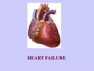
Heart Failure
Heart Failure. PBL 12 April 2011. Question: Define heart failure. Definitions of Heart Failure.


Heart Failure
E N D
Presentation Transcript
Heart Failure PBL 12 April 2011
Definitions of Heart Failure • Harrison’s defines heart failure (HF) as a clinical syndrome that occurs in patients who, because of an inherited or acquired abnormality of cardiac structure and/or function, develop a constellation of clinical symptoms (dyspnoea and fatigue) and signs (oedema and crackles) that lead to frequent hospitalisations, a poor quality of life, and a shortened life expectancy.
Definitions of Heart Failure • Robbins defines congestive heart failure (CHF) as when the heart is unable to pump blood at a rate sufficient to meet the metabolic demands of the tissues or can do so only at an elevated filling pressure. CHF is synonymous with HF.
Question: What is the average weight of the human heart (in males and females)
Definitions of Heart Failure • Heart weight varies with body height and weight, it averages 250-300g in females and 300-350g in males or 0.4-0.5% of body weight. • The free wall of the RV is 0.3-0.5cm, and the LV is 1.3-1.5cm. • Increases in heart weight or ventricular thickness is hypertrophy, enlarged chamber size is dilation, and increased cardiac weight or size from either is cardiomegaly.
Epidemiology of HF • Question: What % of the population suffer from HF? A: 0.1% B: 0.5% C: 1% D: 2% E: 5%
Epidemiology of HF • Answer: 2% • Prevalence rises exponentially with age, affecting 6-10% of people over the age of 65 • Incidence is lower in women than men but women constitute more than 50% of cases because of their longer life expectancy • In North America and Europe, the lifetime risk of developing HF for a 40-year-old is one in five
Global Considerations of HF • What is the major cause of HF in Western countries? • What is the major cause in Africa and Asia in young people? • What is a parasitic cause in South America?
Aetiology of HF • 20-30% of cases of HF with depressed EF have an unknown aetiology. These patients are said to have non-ischaemic, dilated or idiopathic cardiomyopathy.
Hypertrophy • Q: How does cardiac hypertrophy occur? • Q: What are the two broad types of cardiac hypertrophy?
Hypertrophy • Pressure-overload hypertrophy is characterised by concentric increases in wall thickness with new sarcomeres assembled in parallel to the long axes of cells, expanding their cross-sectional area • Volume-overload hypertrophy is characterised by ventricular dilation, with new sarcomeres positioned in series with existing sarcomeres. The wall thickness can be increased, normal or less than normal, so heart weight rather than wall thickness is the best measure of hypertrophy in volume overloaded hearts
Hypertrophy • Heart weights of 2-3x normal are common in patients with systemic hypertension, ischaemic heart disease, aortic stenosis, mitral regurgitation or dilated cardiomyopathy; or 3-4x normal in those with aortic regurgitation or hypertrophic cardiomyopathy • Note that the increased myocyte size is not accompanied with increases in capillary numbers, so the supply of oxygen and nutrients it compromised while oxygen consumption is increased.
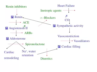
LV Remodelling • LV remodelling develops in response to the mechanical stretch of the myocardium, circulating neurohormones (noradrenaline, ATII), inflammatory cytokines (TNF), other peptides and growth factors (such as endothelin) and reactive oxygen species. • Q: What are the effects of this?
LV Remodelling • Myocyte hypertrophy • Alterations in contractile properties of myocytes • Progressive loss of myocytes through necrosis, apoptosis and autophagic cell death • Β-adrenoceptor desensitisation • Abnormal myocardial energetics and metabolism • Reorganisation of the ECM with dissolution of organised structural collagen and replacement with an interstitial collagen matrix that fails to support myocytes • Expansion of existing infarcts
The LV changes shape from a prolate ellipsoid of revolution to a more spherical shape, which increases meridional wall stress of the LV which creates a de novo mechanical burden for the failing heart • There is increased LV end-diastolic volume and LV wall thinning, creating increased afterload due to LV dilation • High end-diastolic wall stress leads to hypoperfusion of the subendocardium, increased oxidative stress and sustained expression of stretch-activated genes (ATII, endothelin, TNF) • The papillary muscles are pulled apart, resulting in incompetence of the mitral valve and functional mitral regurgitation
Pathology of Left-sided HF HF patients can be categorised as either systolic failure (HF with depressed ejection fraction, or <40%), which has been discussed above, or diastolic failure (HF with preserved EF of >40-50%). Diastolic dysfunction can occur alone or in combination with systolic dysfunction. • Q: What causes left-sided HF? • Q: What are the key morphological features?
Left-sided HF • Left sided HF is most often caused by (1) ischaemic heart disease, (2) hypertension, (3) aortic and mitral valvular diseases and (4) myocardial diseases. • The morphologic and clinical effects of left-sided CHF relate to (1) congestion of pulmonary circulation, (2) stasis of blood in the left heart and (3) hypoperfusion of tissues
Right-sided HF • Q: What is the most common cause? • Q: What is the term given to right-sided HF due to lung pathology, what what are the most common causes of this? • Q: What is the morphology of right-sided HF?
Right-sided HF • Left-sided HF is the most common cause • Cor pulmonale, it is most commonly caused by parenchymal lung disease, other causes include primary pulmonary hypertension, recurrent PE or hypoxia
Morphology of right-sided HF • Heart: Depends on cause (valves, IE), but often hypertrophy and dilation of RA and RV since aetiology is pulmonary • Liver and portal system are congested • Nutmeg liver centrilobular necrosis cardiac sclerosis cardiac cirrhosis • Congestive splenomegaly (size = ?) • Oedematous bowel interferes with absorption • System venous congestion = pleural ( atelectasis), pericardial, peritoneal effusions • Congested kidneys = fluid retention, azotaemia • Pedal, pretibial, sacral oedema anasarca
Prognosis of HF • Development of symptomatic HF still carries a poor prognosis. 30-40% of patients will die within 1 year of diagnosis and 60-70% die within 5 years, mainly from worsening HF or as a sudden event. Patients with symptoms at rest (NYHA class IV) have a 30-70% annual mortality rate whereas patients with symptoms with moderate activity (NYHA class II) have an annual mortality ate of 5-10%, so functional status is an important predictor of patient outcome.
Symptoms of HF • Fatigue, dyspnoea • Orthopnoea (nocturnal cough) • PND (cardiac asthma) • Cheynes-Stokes respiration • Acute pulmonary oedema • Anorexia, nausea, early satiety • Cerebral symptoms (confusion, disorientation, sleep, mood) • Nocturia (insomnia)
Signs of HF • BP is normal or high in early HF but reduced in advanced HF. Pulse pressure may be reduced • Sinus tachycardia (due to NA) • Peripheral vasoconstriction (due to NA) • Elevated JVP • Pulmonary crackles • Pleural effusion • Cardiomegaly, displaces apex beat • S3 if volume overloaded, S4 if diastolic dysfunction, MR and TR if advanced HF • Hepatomegaly, ascites, jaundice • Peripheral oedema • Cardiac cachexia