Heart Failure
Heart Failure. Susan Schayes, MD, MPH Program Director Emory Family Medicine Residency Program Adapted from Dr. Joel Felner and Dr. Eddie Needham. Objectives. Define Heart Failure Know the 5 year mortality rate for heart failure


Heart Failure
E N D
Presentation Transcript
Heart Failure Susan Schayes, MD, MPH Program Director Emory Family Medicine Residency Program Adapted from Dr. Joel Felner and Dr. Eddie Needham
Objectives • Define Heart Failure • Know the 5 year mortality rate for heart failure • Distinguish between New York Heart Association classes (I – IV) and the new American College of Cardiology stages (A – D) • Review and become familiar with treatment options • Know the three beta-blockers demonstrating benefit, and the two that are FDA approved
Objectives • Know indications for an ICD • Know percent of patients who have diastolic dysfunction
Pre-lecture Needs Assessment • What are the four NYHA classes of HF? • What are the four ACC stages of HF? • Which medication classes are routinely prescribed in heart failure? • Which three beta-blockers are approved to treat HF?
DEFINITION • Clinical syndrome • Inability of the heart to produce sufficient cardiac output to meet the metabolic demands of the peripheral tissues while operating at normal filling pressure.
Define Heart Failure • “Heart failure is a complex syndrome that can result from any structural or functional cardiac disorder that impairs the ability of the ventricle to fill with or eject blood.” 1 • The cardinal symptoms are dyspnea and fatigue, while the predominant clinical sign is fluid retention (rales, elevated jugular venous pulsations, and pedal edema). Given that not all patients are volume overloaded at the time of diagnosis (diastolic dysfunction), the term “heart failure” is now preferred over “congestive heart failure.” 1Hunt S, et al, ACC/AHA Guidelines for the Evaluation and Management of Chronic Heart Failure in the Adult: A Report of the American College of Cardiology/American Heart Association Task Force on Practice Guidelines (Committee to Revise the 1995 Guidelines for the Evaluation and Management of Heart Failure). 2001, ACC web site, accessed November 12, 2004.
CLASSIFICATION 1. Acute (pulmonary edema) 2. Chronic stable a. Systolic / Diastolic dysfunction 3. Right / Left ventricular failure 4. High output states
ACUTE PULMONARY EDEMA • Definition:Sudden change: structure/function(LVFP) • Etiology: Cardiac -myocardial (ischemia / infarction) -mechanical (acute regurg; HTN urgency) -electrical (tachycardia: AF/VT) Non-cardiac -high altitude pulmonary edema (HAPE) -heroin overdose; chlorine,etc
Pulmonary edema is caused by (1) imbalance of the Starling forces in the lung (cardiogenic)(2) disruption in the alveolar capillary membrane (non-cardiogenic).
CARDIOGENIC PULMONARY EDEMA NON-CARDIOGENIC PULMONARY EDEMA
1. “hydrostatic APE” Acute cardiogenic or volume-overload pulmonary edema -sudden in pulmonary venous pressure pulmonary interstitial and alveolar fluid -pulmonary and lymphatic drainage can’t compensate acutely to remove the fluid
continued Hallmark: rapid increase in hydrostatic pressure in the pulmonary capillaries causing increased transvascular fluid filtration. It is usually due to pulmonary venous pressure from LVEDP/ LAP. As LAP rises above 25 mmHg fluid breaks thru the lung epithelium flooding the alveoli with protein poor fluid.
2. “-permeability pulmonary edema” (acute lung injury) Non-cardiogenic pulmonary edema -Lymphatic drainage cannot compensate for the lung water caused by the disrupted alveolar capillary membrane. -Caused by vascular permeability of the lung flux of fluid into the interstitium and air spaces
APE with NORMAL HEART SIZE*CARDIAC CAUSES • Acute MR (torn chordae / ruptured PM) • Acute AR (dissection / flail leaflet) • Mitral stenosis • Ischemic HD: AMI / stunned myocardium • Malignant HTN • Acute rapid AF (WPW) *Enlarged heart: Exacerbation of chronic HF; Myocarditis
APE: NON-CARDIAC CAUSESPATHOPHYSIOLOGY • Lung injury damages alveolar-capillary membrane “capillary leak syndrome” ie, transudation of fluid from pulmonary capillaries to alveoli • oncotic pressure (hypoalbuminemia) • Impaired lymphatic drainage
SYSTOLIC DYSFUNCTION • Defect: -myofibrils cannot shorten against a load • Various clinical presentations -asymptomatic, w/ ejection fraction -evidence of CO: fatigue/confused/BUN -evidence of congestion: DOE/leg edema -dilated LV chamber on chest x-ray • Annual mortality -NYHA II-III: 15-20% / NYHA IV: 50%
DIASTOLIC DYSFUNCTION • Pathophysiology: “stiff” ventricle LV: poorly compliant; filling/relaxation -systolic function: normal or markedly -evidence of HF: 35% • Etiology: --ischemia/LVH/fibrosis/normal aging • Symptoms: congestive (pul venous HTN) • Signs: apex-normal/ sustained+S4 • Hemodynamic abn: LVEDP / LAP • Prognosis: not as bad as systolic dysfn
LEFT HEART FAILURE • Etiology -CAD / HTN / Valvular HD / etc • Symptoms -fatigue/congestion (SOB / DOE) • Signs -narrow pulse pressure -hypokinetic carotid pulse -inferolaterally displaced apex -S3/S4 gallops; murmurs of MR/TR
RIGHT HEART FAILURE • Etiology-lung disease: parenchymal / vascular congenital: ASD / Ebstein’s anomaly • Symptoms -fatigue / syncope / girth / edema • Signs -hypotension / parasternal lift distended neck veins / + HJ reflux -right-sided S3 / S4; murmur of TR hepatomegaly / ascites / peripheral edema
HIGH OUTPUT FAILURE Non-cardiac circulatory overload • Etiology -fistula / anemia / pregnancy / hyperT4 • Pathophysiology -SV: preload (VR) + PVR(vasodilate) -CO at rest: afterload / preload -blood volume due to xs Na/H2O • Symptoms: congestion (PCWP) • Signs:HR / SBP/DBP / wide PP / S3
CLINICAL EVALUATION- HF • Risk factors for CAD • Symptoms -only weakly related to LV dysfunction • Fluid status: serum Na / weight / edema • Functional status: NYHA classification
PRECIPITATING FACTORS • Diet: xs Na / H2O; alcohol • Non-compliance with medications • Arrhythmia • Infection • Anemia • Stress • Metabolic: thyroid disease / renal failure
LABORATORY EVALUATION 2-D ECHO / DOPPLER • Most useful test • Determines primary abnormality • Derives Ejection Fraction (EF) -most important single measurement -but, poor correlation with symptoms • Distinguishes systolic / diastolic dysfn • Guide to prognosis (EF and ESV) • Assesses disease progression (remodels)
PATHOPHYSIOLOGY • Ventricular injury / myocyte loss a. Chronic: CAD / HTN / valvular disease b. Acute: AMI / myocarditis / MR / AR • Compensation a. Ventricular remodeling -initially adaptive and benficial -eventually maladaptive and harmful b. Peripheral remodeling • Decompensation
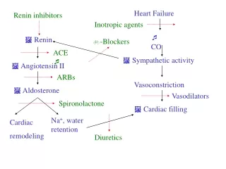
PATHOPHYSIOLOGY: THEORIES • OLD: hemodynamic disorder- ejection (EF) sx (fatigue / dyspnea) -Rx: contractility: inotropes unload periphery: dilators / diuretics • CURRENT: uncontrolled LV remodeling -chamber dilates (spherical); hypertrophy -mechanism: neurohormonal system -Rx: counteract RAAS / SNS • FUTURE: genetic abn / xs cytokines
PATHOPHYSIOLOGY: EVENTS • Primary response: SNS activation (/NE) -initiates vicious circle: afterload • Secondary response: hormone constriction -RAAS: periph perfusion (Na retained) -Vasopressin: non-osmotic release • Vascular endothelial dysfunction (NO) • Result of neurohormonal compensation -adaptive / beneficial: maintains perfusion -long term: maladaptive / deleterious
COMPENSATORY MECHANISMSCOUNTERACTS SV and CO • Starling effect: preload -limited role • muscle (LVH): vs myocyte loss -key • neurohumoral action:contractility-bad -SNS: EPI / NE (HR / PVR) -RAAS: Na/H2O retention;K/Mg;GFR -endothelin / vasopressin / prostacyclin • Brain natriuretic peptide (BNP) -diagnostic / prognostic • Dilatation / remodeling
VENTRICULAR REMODELING • Definition -altered chamber geometry -disproportionate cavity to wall thickness • Pathophysiology-altered extracellular matrix myocfibrosis -up-regulates pro-inflam cytokines -myocyte hypertrophy/apoptosis; -inotropy -imbalance between production of O- / NO -rearranges myocardial fibers: alters length/width ratio
SYSTOLIC DYSFUNCTION DIASTOLIC DYSFUNCTION NORMAL LV Press. Left Ventricular volume LV PRESSURE-VOLUME LOOPS: SYSTOLIC DYSFUNCTION: Contractility: ejection impaired DIASTOLIC DYSFUNCTION: Chamber stiffness: filling impaired
FRANK-STARLING LV FUNCTION CURVES Normal LV Review cardiac physiology to understand these curves Stroke Volume Low CO LV Failure Congestion 10 15 20 LVEDP THE RELATIONSHIP BETWEEN SV and LVEDP
MYOCARDIAL DYSFUNCTION / FAILURE DIASTOLIC DYSFUNCTION SYSTOLIC DYSFUNCTION ENDOTHELIAL DYSFUNCTION LVEDP ARTERIAL BLOOD VOL CO RESERVE NO ENDO- THELIN Periph cap press PCP SNS (NE) VASO- PRESSIN RAA FATIGUE/ RENAL DYSFN Periph constrict Renal constrict ALDO- STERONE PVR ANF CONGESTION Na/H2O retention Vascular stiffness Peripheral Pulmonary EDEMA DYSPNEA PLASMA VOLUME LA cavity
Epidemiology of Heart Failure • Approximately 5 million patients in the USA have HF, with a yearly incidence of close to 500,000. • It is primarily a disease of the elderly, with 6-10% patients over 65 years old being diagnosed with HF. • 80% of hospitalized patients with HF are > 65yo. • Heart failure is the most common Medicare DRG.
Epidemiology of Heart Failure • “…one-year mortality of approximately 45 percent.” 2 • “Survival ranges from 80% at 2 years for patients rendered free of congestion to less than 50% at 6 months for patients with refractory symptoms.” 3 2 Jessup M, Brozena S, Medical Progress: Heart Failure, NEJM, 348(20): 2007-18, 2003. 3 Nohria A, et al, Medical Management of Advanced Heart Failure, JAMA, 287(5): 628-40, 2002.
Epidemiology of Heart Failure • “Heart failure admission rates are rising, and the prognosis of heart failure has been compared with that of malignancy, with a 6-year mortality rate of 84% in men and 77% in women.” 4 • Heart failure kills people much more surely than most cancers! • Coronary artery disease is the cause of two thirds of left ventricular systolic dysfunction 4 Mair F, et al, Evaluation of suspected left ventricular systolic dysfunction, JFP, 51(5): 466-71, 2002.
Diagnosing Heart FailureSymptoms • Decreased exercise tolerance • Fluid retention • Fatigue • Incidentally noted left ventricular dysfunction in an asymptomatic patient
Diagnosing Heart FailureClinical Signs • Elevated jugular venous pressure • Pulmonary rales • S3 • S3 – volume overload • S4 – pressure overload • Peripheral edema
Auscultatory Findings • S3 • S4 • http://www.egeneralmedical.com/listohearmur.html • Rales • http://www.wilkes.med.ucla.edu/intro.html
Diagnosing Heart Failure • Many different terms: • Left vs right-sided failure • Backward vs forward failure • Volume vs pressure overload • Systolic vs diastolic dysfunction – there is a lot of overlap as many patients have aspects of both entities