Download
1 / 49

E N D
DNA Repair M.Prasad Naidu MSc Medical Biochemistry, Ph.D.Research Scholar
Introduction • The maintenance of the integrity of the information in DNA molecules is of utmost importance to the survival of the species . • The major responsibility for the fidelity of replication resides in specific pairing of nucleotide bases . • Proper pairing is dependent upon the presence of favoured tautomers of the purine & pyrimidine nucleotides .
contd • Physiological conditions strongly favors the amino & lactam forms , the unfavored tautomers may participate in mutagenic events if they were unrepaired . • The equilibrium where by one tautomer is more stable than another is only about 104 or 105 in favor of that with great stability. • The favoring of preferred tautomers & the proper base pairing could be ensured by monitoring the base pairing for 2 times .
contd • Double monitoring appear in both mammalian & bacterial systems . • First monitoring occurs at the time of insertion of the deoxyribonucleoside triphosphates , & later by a follow up ,energy requiring mechanism which removes all improper bases that may occur in the newly formed strand . • Unfavored tautomers occur more frequently than once in every 10 8 – 10 10 base pairs .
contd • The mechanisms responsible for DNA repair in E .coli include the 3’ to 5’ exonuclease activities of one of the subunits of polymerase III complex & of the polymerase I molecule . • The analogous mammalian enzymes ( α & δ ) do not posses nuclease proofreading function.
contd • Replication errors occurs even with efficient repair system lead to the accumulation of mutations. • Damage to DNA occurs by environmental , physical & chemical agents classified to 4 types .
The nature of mutations Simple mutations: Transitions(pyrimidine-to-pyrimidine and purine-to-purine) Transversions(pyrimidine-purine and purine-to-pyrimidine) Insertions and deletions (a nucleotide or a small number of nucleotides) ★point mutations: mutations that alter a single nucleotide
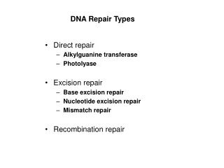
Abnormal regions of DNA , either from copying errors or DNA damage are replaced by 4 mechanisms • Mismatch repair , • Base excision repair , • Nucleotide excision repair , • Double stranded break repair .
Mismatch Repair • Mismatch repair corrects errors made when DNA is copied , for example a Cytosine could be inserted opposite an A , or the polymerase could slip or stutter & insert 2 – 5 extra unpaired bases . • Specific proteins scan the newly synthesized DNA , using adenine methylation within GATC sequence as the point of reference .
contd • The template strand is methylated & newly synthesized strand is not methylated . • This difference allows the repair enzymes to identify the strand that contains the errant nucleotide which requires replacement . • If a mismatch or small loop is found , a GATC endonuclease cuts the strand bearing the mutation at a site corresponding to the GATC .
contd • An exonuclease digests this strand from GATC through the mutation thus removing the faulty DNA . • The above digestion can occur from either side if the defect is bracketed by 2 GATC sites . • The defect is filled by normal cellular enzymes according to the base pairing rules.
In E .coli three proteins ( Mut S , Mut L & Mut H ) are rrequired for recognition of the mutation & nicking of the strand . Other cellular enzymes ligase , polymerase & SSBs remove & replace the strand .
MutS scans the DNA, & recognize the mismatch or the distortion in the DNA backbone .
Clinical importance • Faulty mismatch repair is linked to hereditary nonpolyposis colon cancer ( HNPCC ) . • Genetic studies linked HNPCC in some families to a region of chromosome 2 . • The gene on chromosome 2 is hMSH2 is human analogue of Mut S protein that is involved in mismatch repair . • Mutations of hMSH2 account for 50 - 60 % of HNPCC .
contd • Another gene hMLH1 is associated with most other cases . • hMLH1 gene is human analogue of bacterial mismatch repair gene Mut L . • Microsatellites are repeated sequences of DNA. • These repeated sequences are common, and normal. • The most common microsatellite in the humans is a dinucleotide repeat of CA, which occurs tens of thousands of times across the genome .
contd • Muted hMSH2 & hMLH1 mismatch repair enzymes results in increased size of microsatellites , this must affect the function of a protein critical in surveillance of the cell cycle in these colon cells . • The appearance of abnormally long or short microsatellites in an individual's DNA is referred to as microsatellite instability. • Microsatellite instability (MSI) is a condition manifested by damaged DNA due to defects in the normal DNA repair process.
Base Excision Repair • This mechanism is suitable for replacement of a single base but is not effective at replacing regions of damaged DNA . • The depurination of DNA which happens spontaneously due to the thermal lability of the purine N – glycosidic bond , occurs at a rate of 5000 – 10,000 /cell / day at 37 ° C .
contd • Cytosine , adenine & Guanine bases in DNA spontaneously form uracil , hypoxanthine or xanthine respectively . • None of the above are normal bases . • N – glycosylases can recognize these abnormal bases & remove the base itself from the DNA . • This removal marks the site of the defect & allows an apurinic or apyimidinic endonuclease to excise the abasic sugar .
contd • The proper base is replaced by repair , DNA polymerase & the ligase returns the DNA to its original state , this series of events is called base excision repair . • By similar series of steps involving initially the recognition of the defect , alkylated bases & base analogues can be removed from DNA .
Deamination C-U Depurination ----> an abasic site Deamination of 5-mC---->T
DNA is damaged by Alkylation, Oxidation, and Radiation Often mispair with thymine G:C –A:T Reactive oxygen species O2-, H2O2, OH• G modification (alkylation & oxidation)
Mutations are also caused by base analogs and intercalating agents Base analogues
Base excision repair pathway (apurinic/apyrimidinic; recognizes missing base)
Nucleotide Excision Repair • This mechanism is used to replace regions of damaged DNA up to 30 bases in length . • UV light induces the formation of cyclobutane pyrimidine – pyrimidine dimers . • Smoking causes formation of benzopyrene – guainine adducts .
Incapable of base-pairing and cause the DNA polymerse to stop during replication Thymine dimer by ultraviolet light
contd • Ionizing radiation , cancer chemotherapy & chemicals found in environment cause base modification , strand breaks , cross – linkage between bases on opposite strand or between DNA protein & numerous other defects are repaired by this mechanism . • Nucleotide excision repair is complex process involves more gene products than 2 other types of repair , essentially involves hydrolysis of 2 phosphodiester bonds on the strand containing the defect .
contd • A special excision nuclease ( exinuclease ) consisting of at least 3 sub units in E .coli & 16 polypeptides in humans . • In eukaryotic cells the enzymes cut between the 3rd to 5th phosphodiester bond 3 ‘ from the lesion & on the 5’ side the cut is some where between the 21st & 25th bond . • Thus a fragment of 27 – 29 nucleotides long is exicised . • After the strand is removed it is replaced by exact base pairing through the action of polymerase ( δ/ε in humans), ends are joined by DNA ligase.
2 3 1 4
1.UvrA and UvrB scan DNA to identify a distortion 2. UvrA leaves the complex,and UvrB melts DNA locally round the distortion 3. UvrC forms a complex with UvrB and creates nicks to the 5’ side of the lesion 4. DNA helicase UvrD releases the single stranded fragment from the duplex, and DNA Pol I and ligase repair and seal the gap
Transcription coupled DNA repair: nucleotide excision repair system is capable of rescuing RNA polymerase that has been arrested by the presence of lesions in the DNA template
Clinical Imporatance • Xeroderma pigmentosum is an autosomal recessive genetic disease . • The clinical syndrome include marked sensitivity to sunlight ( UV rays ) with subsequent formation of multiple skin cancers & premature death . • The risk of developing skin cancer is increased 1000 to 2000 fold .
contd • The inherent defect seems to involve the repair of damaged DNA , particularly thymine dimers . • Cells cultured from patients with xeroderma pigmentosum exhibit low activity for the nucleotide excision repair process . • Seven complementation groups have been identified using hybrid cell analysis so at least 7 gene products ( XPA – XPAG ) .
contd • XPA & XPC are involved in recognition & excision .XPB & XPD are helicases & interestingly are subunits of the transcription factor TFIIH .
Double Strand Break Repair • The repair of double strand breaks is part of the physiological process of immunoglobulin gene rearrangement . • It is also important mechanism for repairing damaged DNA such as occurs as result of ionizing radiation or oxidative free radical generation . • Some chemotherapeutic agents destroy cells by causing double stranded breaks or preventing their repair .
contd • Two proteins are involved in the nonhomologous rejoining of a ds break . • Ku , a hetero dimer of 70 & 86 kDa subunits , bind to free DNA ends & has latent ATP dependent helicase activity . • The DNA bound Ku hetero dimer recruits an unusual DNA dependent Protein kinase ( DNA – PK )
contd • DNA – PK has a binding site for DNA free ends & another for ds DNA just inside these ends . • It allows the approximation of the 2 separated ends . • The free end DNA/Ku/DNA – PK complex activates the kinase activity in the later . • DNA – PK reciprocally phosphorylates Ku & the other DNA – PK molecule on the opposing strand , in trans .
contd • DNA – PK then dissociates from the DNA & Ku, resulting in activation of the Ku helicase. • This results in unwinding of the 2 ends . • The unwound approximated DNA forms base pairs . • The extra nucleotide tails are removed by an exonuclease & the gaps are filled and closed by DNA ligase .
Some repair enzymes are multifunctional • DNA repair proteins can serve other purposes example some repair enzymes found as components of the large TFIIH complex that play a central role in gene transcription . • Another component of TFIIH is involved in cell cycle regulation . • Thus three critical cellular processes may be linked through use of common proteins .
Clinical importance • In patients with ataxia telangiectasia ,an autosomal recessive disease characterized by cerebellar ataxia & lymphoreticular neoplasms , in these patients there appears to exist an increased sensitivity to damage by X rays . • Fanconis anemia an autosomal recessive anemia characterized by an increased frequency of cancer & by chromosomal instability , probably have defective repair of cross linking damage.