Download
1 / 13
140 likes | 332 Views
Cellular structure of nervous system.

E N D
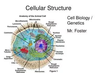
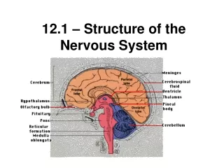
Cellular structure of nervous system The human nervous system contains at least 10 billion neurons. These basic building blocks of the nervous system have evolved from primitive neuro-effector cells that respond to various stimuli by contracting . nerve tissue is distributed throughout the body as an integrated communications network. Anatomically ,the nervous system is divided in to:- 1- the central nervous system which it consist of the brain and spinal cord 2- the peripheral nervous system ,composed of nerve fibers and small aggregates of nerve cells. Structurally, nervous tissue(system) consist of 2 classes of cell types :- 1- Nerve cells ,or nurons, which usually show numerous long processes. 2- several types of glial cells , or neuroglia , which support and protect neurons and participate in neural activity ,neural nutrition, and the defense processes of the central nervous system.
Neurons respond to environmental changes (stimuli) by altering electrical potential differences that exist between the inner and outer membranes . • cells with this property : neurons ,muscle cells , some gland cells are called excitable or irritable . • Neurons react promptly to stimuli the modification of electrical potential may be restricted to the place that rereceived the stimulus or may spread propagated throughout the neuron by the membrane . this propagation called the action potential or nerve impulse which transmits information to other neurons ,muscles or gland • Na+ Na+ Na+ Na+ Na+ Na+ • ____________________ • K+k+ k+ k+ k + k+ k+ k+ k+
Neuron structure • Although neurons vary in size and shape , they have certain features in common, these include cell body (perikaryon or soma ) and cytoplasmic processes extensions or nerve fibers. Which conduct impulses to ,or from the cell body . the perikaryon cotntains:- • amass of granular cytoplasm ,cell membrane ,numerous mitochondria ,lysosomes golgi apparatus and a net work of fine threads called neurofibrils which extend in to nerve fibers. • Near the center of the cell body there is alarge spherical nucleus with prominent nucleolus. This nucleus does not undergo mitosis and consequently mature neurons seems .to be incapable to reproduction.The cytoplasm of neurons has agranular material that stain intenselywith basic dyes these granules are known as Nissl bodies which represent sites of active protein synthesis . • Two kinds of processes arise from the cell body , ashorter processes branching called dendrites and one long process called Axon.
Dendrites • Most nerve cells have numerous dendrites which increase considrably the receptive area of the cell .dendrites arborization (branching) makes it possible for one neuron to receive and integrate with a great number of axon terminals from other nerve cells. • the composition of dendrites cytoplasm is very similar to that of the perikaryon , however dendrites are devoid (without ) golgi complex . Nissl bodies and mitochondria are present except in very thin dendrites . • usually its short and divide like the braches of a tree and covered by a large number of thorny spines which are small dendritic projections representing sites of synaptic contact.
Axons • Most neurons have only one axon ; a very few do not have an axon at all. An axon is a cylindrical process that varies in length and diameter according to the type of neuron. • all axon originate from short pyramid shaped region called the axon hillock which usually arises from the perikaryon but in a few cases originates from the stem of amajor dendrites , the axon hillock can be differentiated from dendrites by distinctive cytologic features. • the rough endoplasmic reticulum and ribosomes found in perikaryons and dendrites but do not extend in the axon hillock . • In the axon hillock , the microtubules are arranged in fascicles or bundles . the plasma membrane of the axon is called the axolema and its contents called axoplasm
Types of neurons • Neurons vary considerably in the size and shape of their cell bodies and in the length and manner of branching of their processes .according to these factors the neurons are classified in to :- • 1-Unipolar neurons :- • A neuron with single process arising from the cell body .it is rare in adult vertebrates. • 2- Pseudounipolar neuron :- • which has process that bifurcates , one branch passing peripherally and as dendrites .and other passing centrally as axon such as the neuron in the cranial and spinal ganglia . • 3-Bipolar neurons:- it is un common also it has single axon and single dendrites ,usually located at apposite sides of spindle shaped soma,it is found in retina, olfactory epithelium. • 4- Multipolar neurons:- • with numerous dendrites and usually single axon ,preseeent in autonomic ganglia.
According to the functional differences it can be grouped s follows:- • Sensory neurons:-,are those that carry nerve impulse from peripheral body parts 9skin) in the brain or spinal cord . • Inter neurons : or association neurons, lie within C.N.S.. They form links between other neurons .Its function to transmit impulses from one part of the brain or spinal cord to another. • Motor neurons: or efferent neurons ,carry nerve impulses out from C.N.S. to effectors ( parts of the body such as muscles or glands ).
Types of nerves Types of nerves • Sensory nerves or afferent nerves that conduct impulses in to C.N.S. • Motor or efferent nerves that carry impulses out , to the muscles or glands (effectors) most nerves include both types, they are so called
Neuroglia • Neuroglia :- are supporting cells of C.N.S. , there are several types and more numerous than neurons .they fill spaces .suppor neurons and generally bind together in the nervous tissue of C.N.S. • Neuroglial cells are smaller than neurons , some of glial cells are smaller than neurons ,some of glial cells mobile and ,unlike neurons ,retainthe ability to divide. • Neurogllial cells may be divided in to two major categories. • Macroglia: or large glial cells. • Microglia : or small glial cells. • Macroglial cells are derived from ectoderm of the neural tube , in contrast, • Microglial cells are derived from the mesoderm.
Types of neuroglia • Astrocytes ( macroglial cell ): • are star –shaped cells have many radiating cytoplasmic processes or extensions with expanded tips called astrocyte pedicles or feed that adhere both to capillaries and to neurons . • Astrocytes have large ovoid or spherical nucleus, the cytoplasm contains golgi complex, few ribosomes ,lysosomes and glycogen,and there are bundles of glial filaments similar to neuro filaments. • Two types of astrocytes are recognized: - • Protoplasmic astrocytes : seen mainly in gray matter of C.N.S., they have short ,thick processes with many branches. • Fibrous astrocytes : are found mainly in white matter and have only a few thin and large processes , with few or no branches.the main functions are supporting the C.N.S. and involve in repair mechanism after brain damage .
2- Oligodendrocyt(macroglial cells) • Are smaller than astrocytes with fewer shorter cell processes ,it has small and dar4k nuclei and scanty cytoplasm with numerous free and attached ribosomes ,an extensive golgi apparatus and many mitochondria and microtubules. • They are found in two places :- • in the gray matter near the cell bodies of neurons • among bundles of axons in white matter. • Oligodendrocytes function in the formation of myelin within the C.N.S. ;thus serving the same function as schwann cells in the P.N.S.. however un like schwann cells each oligodendrocytes has several processes and these form myelin sheath,around several adjacent nerve fibers.
3-Glioblast (microglia cells) • These are smallest neuroglial cell. The cell body is flattened with short spiny processes few in number and scattered throughout the C.N.S. • if THE C. BN.S. damaged they increase in size and become actively motile and phagocytic.
Ependymal cells • They are supporting cells lining the ventricles of the brain and central canal of spinal cord , where they constitute continuous epithelial lining known as the ependyma. these cells are cuboidal to low columnar in shape and have few cilia on their free surface and they cover specialized parts within the brain (choroids plexus).