Download
1 / 25
250 likes | 276 Views
This article explores the structure and anatomy of the nervous system, including the neuraxis, anatomical directions, planes of section, two nervous systems, meninges, cerebrospinal fluid, brain development, cerebral cortex, primary sensory and motor cortex, four lobes and association cortex, limbic system, basal ganglia, diencephalon, mesencephalon, metencephalon, and myelencephalon.

E N D
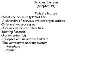
Biological Bases of Behavior 3: Structure of the Nervous System
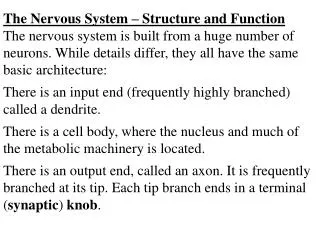
Neuroanatomy Terms • The neuraxis is an imaginary line drawn through the spinal cord up to the front of the brain • Anatomical directions are understood relative to the neuraxis • Anterior (rostral): toward the head • Posterior (caudal): toward the tail • Ventral (inferior): toward the “belly” • Dorsal (superior): toward the back (top of head) • Location in brain: • Ipsilateral: same side of brain • Contralateral: opposite side of brain 3.2
Planes of Section • The brain can be sectioned in three planes • Each section provides a different view of the internal anatomy of the brain • Sagittal • Coronal (or transverse) • Horizontal 3.4
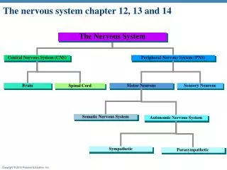
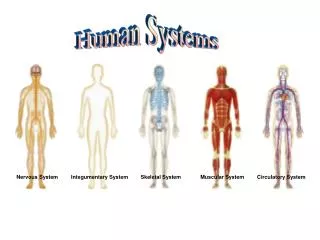
Two Nervous Systems • The nervous system consists of two divisions • The central nervous system (CNS) is comprised of the brain and spinal cord • Spinal cord is a conduit for information to and from brain • The peripheral nervous system (PNS) is comprised of the cranial/spinal nerves and peripheral ganglia • PNS nerves project to target organs and to muscles (efferent) • These nerves also carry sensory information to the brain (afferent) 3.5
The Meninges • CNS are protected by a series of membranes termed meninges • Dura mater-outer (thick) layer • Arachnoid-middle layer • Arachnoid membrane • subarachnoid space filled with cerebrospinal fluid (CSF) • Major blood vessels run through the arachnoid layer • Pia mater- inner layer • Overlies every detail of the outer brain, smaller blood vessels run through • PNS are protected by fused dura and pia membrane Source: Brain Tumor Foundation of Canada. http://www.btfc.org/ 3.6
Cerebrospinal Fluid • The brain floats in a pool of cerebrospinal fluid (CSF) which reduces its net weight from 1400 g --> 80 g • CSF is also contained within four brain ventricles • CSF is produced by the choroid plexus of each ventricle • The brain ventricles are an access point for drug studies • The brain ventricles can expand when brain cells are lost (as in alcoholism or certain diseases) 3.7
Brain Development • The nervous system develops from ectoderm (outer layer) which forms a plate (~day 18) • The edges of the plate curl and eventually fuse together forming a neural tube • By ~day 28, the rostral end of the neural tube has formed the ventricles and the tissue that surrounds these hollow chambers has formed three major divisions of the brain • Forebrain, midbrain, and hindbrain 3.8
Overview of the CNS Spinal cord 3.10
Cerebral Cortex • The cerebral cortex forms the outer surface of the cerebral hemispheres • Cortex surface is convoluted by grooves • Sulci (small grooves) • Fissures (large grooves) • The bulges in cortex are termed gyri • The cortex is primarily composed of cells, giving it a gray appearance • The cortex is formed from 6 layers of cells • Cortex can be divided into 4 lobes: frontal, parietal, occipital, and temporal 3.11
Limbic System • The limbic system is comprised of • Hippocampus: involved in learning and memory • Amygdala: involved in emotion • Mammillary Bodies • The fornix is a fiber bundle that interconnects the hippocampus with the mammillary bodies • Limbic cortex 3.14
Basal Ganglia • The basal ganglia are a collection of subcortical nuclei that lie just under the anterior aspect of the lateral ventricles • “Ganglia” is a misnomer (term refers to collections of cell bodies in periphery) • Basal ganglia consist of: • Globus pallidus, Caudate nucleus, Putamen • Basal ganglia are involved in the control of movement • Associated with Subthalamic nucleus, Substantia nigra 3.15
Diencephalon • Diencephalon consists of • Thalamus: contains nuclei that receive sensory information and transmit it to cortex • Hypothalamus: contains nuclei involved in integration of species-typical behaviors, control of the autonomic nervous system and pituitary 3.16
Mesencephalon • The mesencephalon (midbrain) consists of • Tectum is the dorsal portion of midbrain • Superior colliculi: visual system • Inferior colliculi: auditory system • Tegmentum is the portion of the midbrain located under the tectum and consists of the • Rostral end of the reticular formation • Periaqueductal gray • Red nucleus • Substantia nigra • Ventral tegmental area 3.17
Metencephalon • Metencephalon consists of the • Pons • Contains the core of the reticular formation • The pons is involved in the control of sleep and arousal • Cerebellum is involved in motor control 3.18
Myelencephalon • The myelencephalon consists of the • Medulla oblongata • The medulla is the most caudal portion of brain and is rostral to the spinal cord • The medulla contains part of the reticular formation • The nuclei of the medulla control vital functions such as regulation of the cardiovascular system, breathing, and skeletal muscle tone 3.19
The Spinal Cord 3.20
The Peripheral Nervous System • Somatic division of PNS is comprised by nerves that control muscle action and that carry sensory information back to the CNS • Cranial nerves (12) • Spinal nerves (31) • Autonomic division of PNS governs smooth muscle and gland secretion • Parasympathetic: supports activities that increase energy • Sympathetic: arousal and the expenditure of energy 3.21
Definitions • Nerve: collection of axons outside CNS • Tract: collection of axons inside CNS • Nucleus: collection of cell bodies inside CNS • Ganglion: collection of cell bodies outside CNS 3.22
Somatic Nervous System • Cranial Nerves (12) • Motor only (red), sensory only (blue), mixed function • Spinal Nerves (31 pair) • 8 Cervical • 12 Thoracic • 5 Lumbar • 5 Sacral • 1 Coccygeal 3.23
The Autonomic Nervous System • Sympathetic division • Associated with energy expenditure • Derives from thoracic and columbar levels of the spinal cord • Parasympathetic division • Associated with energy conservation • Derives from cranial and sacral levels of the spinal cord 3.24
Overview of the ANS 3.25