Download
1 / 11
110 likes | 248 Views
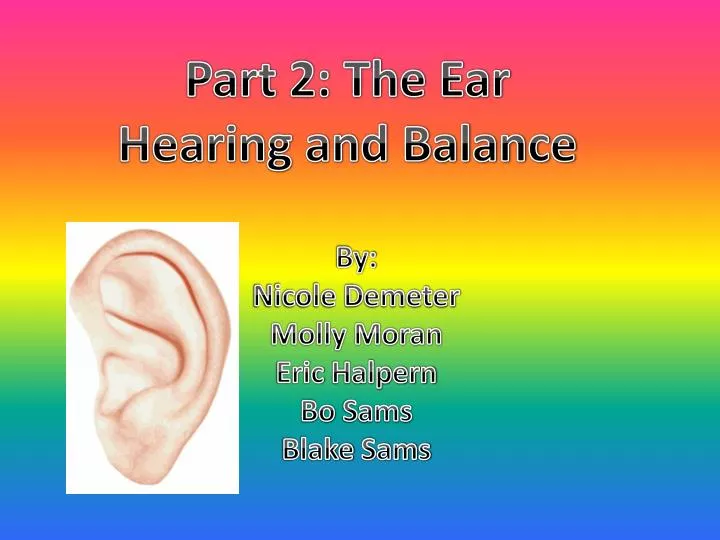
Part 2: The Ear Hearing and Balance. By: Nicole Demeter Molly Moran Eric Halpern Bo Sams Blake Sams. Anatomy of the Ear . External (outer) Ear - is composed of the auricle and the external acoustic meatus . Auricle - shell-shaped structure surrounding the auditory canal opening

E N D
Part 2: The Ear Hearing and Balance By: Nicole Demeter Molly Moran Eric Halpern Bo Sams Blake Sams
Anatomy of the Ear External (outer) Ear- is composed of the auricle and the external acoustic meatus. • Auricle- shell-shaped structure surrounding the auditory canal opening • External acoustic meatus- short, narrow chamber carved into the temporal bone of the skull -Inside the external acoustic meatus are ceruminous glands that produce earwax (cerumen) that traps foreign bodies and protects from insects. -When sound waves move through the auditory canal eventually hitting the tympanic membrane (eardrum), causing it to vibrate.
Anatomy of the Ear Cont. Middle Ear-small, air-filled, mucosa-lined cavity within the temporal bone. It has two openings in the bony wall called the oval window and the round window. Inside the middle ear the pharyngotymphatic tube runs down to connect the ears with the throat. Also, inside the middle ear are the three smallest bones in the body called the auditory ossicleswhich transmit the vibratory motion of the eardrum to the fluids of the inner ear. When the eardrum moves, the hammer (malleus) vibrates the anvil (incus), then vibrates the stirrup (stapes) which presses the oval window, setting the inner ear fluids into motion, exciting the hearing receptors.
Anatomy of the Ear Cont. Internal (inner) Ear-is a maze of bony chambers called the osseous labyrinth, located deep within the temporal bone behind the eye socket. There are three subdivisions of the inner ear. • Cochlea • Vestibule • Semicircular canals - The bony labyrinth is filled with a plasmalike fluid called perilymph. Suspended in the perilymph is a membranous labyrnthcontaining a thicker fluid called endolymph.
Mechanisms of Equilibrium • Vestibular apparatus-equilibrium receptors of the inner ear, divided into static equilibrium and dynamic equilibrium Static Equilibrium- our sense of changes in the position of the head. What causes this sense are receptors called maculae that are inside the vestibule. Each maculae has receptor hairs embedded in the otolithic hair membrane, and covered with stones made of calcium called otoliths. When the head moves, gravity pulls the otolithic membrane, bending the hair cells. This message is sent to the vestibular nerve, telling us the position of our head.
Mechanisms of Equilibrium Dynamic Equilibrium- respond to angular or rotatory movements. The receptors called cristaampullarisare found at the base of the semicircular canal. Cristaampullaris is made of hair cells covered with cupula (gelatinous cap) and as your body moves in circles, the endolymph lags behind your motion to stimulate hair cells. The impulses are then sent to the vestibular nerve.
Mechanisms of Hearing Inside the cochlear duct, is the spiral organ of cortiwhich contains hearing receptors, or hair cells. As the sound wave travels through the middle and inner ear, pressure waves set vibrations in the basilar membrane, which moves the hair cells set in tectoral membrane. Once stimulated, the hair cells transmit impulses along the cochlear nerve, telling our brains about the sounds we hear.
Diseases and Disorders: http://www.google.com/imgres?q=otosclerosis&um=1&hl=en&sa=N&biw=1280&bih=842&tbm=isch&tbnid=nuFG-Qyb3tsLWM:&imgrefurl=http://www.hearingcentral.com/diseasesanddisorders.asp&docid=yKLaK63YFVyrDM&imgurl=http://www.hearingcentral.com/images/otosclerosis.jpg&w=265&h=196&ei=JZvKTs-pAoicgQf2rKBd&zoom=1&iact=rc&dur=172&sig=105948829056681227318&page=1&tbnh=134&tbnw=181&start=0&ndsp=24&ved=1t:429,r:8,s:0&tx=157&ty=61
Diseases and Disorders: treatment for sensorineural deafness http://www.google.com/imgres?q=cochlear+implant&um=1&hl=en&biw=1280&bih=842&tbm=isch&tbnid=P198awrYWUkRpM:&imgrefurl=http://kidshealth.org/parent/general/eyes/cochlear.html&docid=9Mdg2sNMjQfbVM&imgurl=http://kidshealth.org/parent/general/eyes/images_80941/P_cochlear-noConsole.jpg&w=420&h=315&ei=b6DKTpHOEIX7ggex_dlk&zoom=1&iact=hc&vpx=599&vpy=187&dur=2751&hovh=194&hovw=259&tx=102&ty=103&sig=105948829056681227318&page=1&tbnh=142&tbnw=171&start=0&ndsp=23&ved=1t:429,r:3,s:0
Diseases and Disorders: Meniere’s syndrome http://en.wikipedia.org/wiki/M%C3%A9ni%C3%A8re's_disease