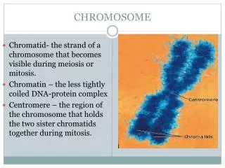
Chromosome disorders
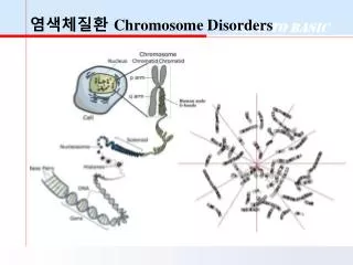
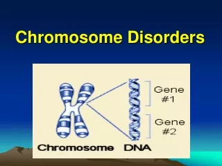
Chromosome disorders. Chromosome disorders are the result of missing or extra chromosomes . Egg and sperm cells (gametes) fuse during Fertilization (syngamy). Types of chromosome disorders. Missing a chromosome = Monosomy – a person inherits only one chromosome of a homologous pair

Chromosome disorders
E N D
Presentation Transcript
Chromosome disorders are the result of missing or extra chromosomes.
Egg and sperm cells (gametes) fuse during Fertilization (syngamy).
Types of chromosome disorders • Missing a chromosome = • Monosomy – a person inherits only one chromosome of a homologous pair • An Extra Chromosome = • Trisomy – a person inherits one extra chromosome of a homologous pair
Nondisjunction: Chromosome disorders are caused by nondisjunction. Nondisjunction occurs during meiosis. Chromosomes fail to separate properly so that gametes have abnormal numbers of chromosomes.
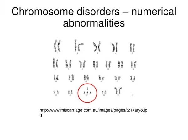
Trisomy Example: Down Syndrome The most common form of trisomy is “trisomy-21.” An extra chromosome is inherited at autosome pair #21. The disorder caused by trisomy 21 is Down Syndrome.
People with Down syndrome can lead active, happy lives. Women who become pregnant later in life, are more prone to have Down syndrome babies. • Women in their 20s who become pregnant have about a one-in-1,230 chance of having a pregnancy affected with Down syndrome. • At age 30, it's one in 690. • By age 35, the chances increase to one in 270. • At age 40, the risk in one in 78. • At age 45, chances are one in 22. They may have fetal cells screened to find out the chromosomes present.(prenatal test)
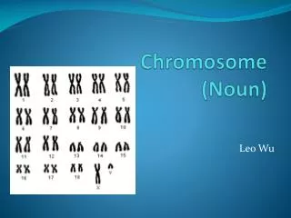
The diagnostic picture of chromosomes is known as a karyotype.
How do doctors get a sample of the fetal cells to make a karyotype? Amniocentesis – Amniotic fluid that surrounds the fetus is drawn into a syringe. It contains some of the fetal cells. Living cells are removed from the amniotic fluid. These cells are then cultured in a medium in which they undergo mitosis.
Amniocentesis - continued Mitosis is stopped in metaphase using chemicals. The cells are then placed onto a slide and spread out. They are viewed under a microscope which is specially adapted with a camera to take a picture of the chromosomes from one cell. A magnified picture is taken of the chromosomes.
Chromosomes on a photograph are cut out and arranged in pairs. This is now done with computer software.
Other Chromosome Disorders:People usually have two sex chromosomes.
Sometimes there can be extra or missing sex chromosomes • Turner’s syndrome(only one X chromosome) X0 • Females who have Turner’s syndrome are often mentally retarded and sterile because their sex organs do not develop. (Some Turners have above average intelligence.) • Klinefelter’s syndrome Males with and extra X chromosome. They are often mentally retarded and sterile. XXY
XYY Syndrome • Boys with XYY syndrome tend to be tall and have difficulties with language. The intelligence quotient (IQ) tends to be slightly lower than that of other family members. Learning disabilities, hyperactivity, attention deficit disorder, and minor behavioral disorders can develop. • The XYY syndrome was once thought to cause aggressive or violent criminal behavior, but this theory has been disproved.
Some Additional Chromosome Disorders
Patau syndrome – (Trisomy 13) The baby may be markedly retarded. Their may be a sloping forehead, a harelip, or a cleft palate. In some cases microcephaly occurs. Polydactyl hands and feet are present (extra fingers and toes). Death may occur in hours or days from organ defects, although some have lesser complications and survive into young adulthood and later (oldest living born 1982.)
Edward syndrome – (Trisomy 18) Babies who have Edward’s syndrome nearly always have problems with their heart, lungs and digestive system. Typical characteristics are a small head, a flat forehead and receding chin. Clenched fingers, a harelip and cleft palate may also be present. It is a rare condition affecting about one baby in 5,000. Death usually occurs in 3 – 4 months.