Download
1 / 37
430 likes | 1.03k Views
Lecture 1 DNA damage. Damage Reversal. Base excision repair. Mismatch repair Lecture 2 Nucleotide excision repair: cellular and clinical aspects Nucleotide excision repair: genes and proteins Lecture 3 Replication of damaged DNA. Mutagenesis and carcinogenesis. Course Learning objectives.

E N D
Lecture 1 DNA damage. Damage Reversal. Base excision repair. Mismatch repair Lecture 2 Nucleotide excision repair: cellular and clinical aspects Nucleotide excision repair: genes and proteins Lecture 3 Replication of damaged DNA. Mutagenesis and carcinogenesis
CourseLearning objectives • To gain an understanding of the molecular mechanisms that maintain genome stability • To appreciate the importance of this topic for human health. Learning outcomes (Lecture 1a) • Understanding: • Different types of DNA damage • Three examples of ways in which cells can reverse damage in situ • Basic mechanism of Base Excision Repair
Procarcinogen Inactive Products Detoxification Ultimate Carcinogen DNA DNA Damage DNA Repair Cell cycle arrest Cell Death Chromosome Rearrangements Mutations Activation of Oncogenes Inactivation of Tumour Suppressor Genes Other disorders Pre-cancerous cells Cancer 1.1 Defence Mechanisms
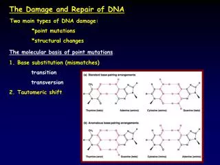
O O O O CH3 O CH3 CH3 1-methyladenine 3-methyladenine O-6-methylguanine DNA Damage N O Major UV photoproducts 1 2 6 H N 2 3 3 1 N 5 4 N 6 5 4 H3C H3C NH2 H 6 4 5 O N 1 N 2 3 N N O H3C TC (6-4) photoproduct (6-4PP) Cyclobutane pyrimidine dimer (CPD) Methylated purines CH3 CH3 4 3 5 2 6 O 1 7-methylguanine 3-methylcytosine 1.2
Aspects of DNA repair 1. Initial damage 2. Repair of damage 3. Genes involved 4. Mechanism of action of gene products 5. Replication of unremoved damage. Cell cycle progression. 6. Biological consequences of damage, repair and failure to repair. 1.3
PR light Survival No PR light UV Dose Cyclobutane pyrimidine dimers Time Damage reversal 1. Photoreactivation Friedberg et al, 2005DNA Repair and Mutagenesis 1.4
Photolyase mechanism Friedberg et al, 2005DNA Repair and Mutagenesis 1.5
O-6-methylguanine CH3 Thymine CH3 CH3 O O-6-methylguanine Damage reversal 2. Repair of O6-methylguanine Methylated purines 6 6 Mispairing of O-6-methylguanine with thymine 1.6
Damage reversal 2. Repair of O6-methylguanine Adaptation Treat E.coli with indicated dose of MNNG, then expose to high dose Control Adapted Friedberg et al, 2005DNA Repair and Mutagenesis 1.7
Dual activities of Ada methyltransferase Friedberg et al, 2005DNA Repair and Mutagenesis 1.8
Induction of ada gene Ogt gene is not inducible Friedberg et al, 2005DNA Repair and Mutagenesis 1.9
Alkyltransferases in mammalian cells • Similar mechanism to E. coli, but for O-6-meG alone, like Ogt, not inducible. • K/o mouse constructed, very sensitive to carcinogenesis by methylating agents. • Conversely transgenic mice bearing MGMT gene are more resistant. • Many cancer cell lines are Mex-. MGMT silenced by methylation in about 50% of tumours. • Mex- cells are sensitive to killing and mutagenesis by alkylating agents. • Many cancer therapy drugs are alkylating agents, eg temozolomide. • Patrin2 binds MGMT and depletes it. Currently in clinical trials together with temozolomide. 1.10
Damage reversal3. Oxidative demethylation (A3) Mechanism ar alkB survival wt alkB Friedberg et al, 2005DNA Repair and Mutagenesis 1.11
Deamination of bases 8-hydroxyguanine
Base Excision-Repair
3-d structures of APendonucleases APE1 ENDO IV
Summary (Lecture 1a) • DNA damage can cause distortions of different severity • UV damage is repaired by photoreversal (not in placental mammals) • O6-methylguanine is repaired by a specific methyltransferase • 1-methyladenine and 3-methylcytosine are repaired by oxidative demethylation • Spontaneous lesions are removed by Base Excision Repair
Learning outcomes (Lecture 1b) • Understanding: • Detailed mechanism of mismatch repair in E. coli and eukaryotes • How mismatch repair is important both for cancer protection and cancer therapy
Mismatch Repair (A5, A6) • DNA polymerases replicate DNA very faithfullyAccurate insertionAssociated 3’-5’ exonuclease for proof-readingError rates c. 10-6 or less • But genomes are big: E. coli 3x10⁶ bp, mammals 3x10⁹ • Errors can be single base mismatches or small insertions or deletions caused by base slippage • Mismatches are repaired by the MMR system which recognises the mismatched bases • But there’s a problem 1.12
Methylation-directed mismatch repair Dam protein Friedberg et al, 2005DNA Repair and Mutagenesis 1.13 Dam- strains (methylation deficient) are mutators
Mismatch recognition and strand discrimination in E. coli MutH, MutL and MutS- strains are mutators CH3 CH3 S2 H L2 1.14
Activities of MutS Sliding clamp Crucial Phe in Phe-X-Glu contacts mismatch Jiricny, NRMCB, 2006 3-D structure of MutS Structural homodimer Functional heterodimer Jiricny, Current Biology, 2000 1.15
Late steps in MMR in E. coli UvrD helicase II unwinds SSB covers exposed ss DNA DNA Ligase 1.16 Friedberg et al, 2005DNA Repair and Mutagenesis
Eukaryotic homologues of MutH,L,S MutL: Mlh1 MMR Mlh2 ? Mlh3 MMR Pms1 ? Pms2 MMR (= Pms1 in yeast) MutS: Msh2 MMR Msh3 MMR Msh4 Meiosis Msh5 Meiosis Msh6 MMR MutH: No homologues Neither yeast nor Drosophila has methylated DNA Strand discrimination based on nicks/ends in daughter DNA MMR proteins interact with PCNA at replication fork 1.17
IDL (Insertion-deletion loop) G C Mismatch Repair in eukaryotes GT G GTC T GT GTAC T Primary recognition MutSa Preference for single base mismatches and small IDL MutSb Preference for large IDL GTC G T Msh2 Msh2 Msh6 Msh3 Secondary recognition MutLa GTC G PCNA PCNA Msh2 T Msh6 Msh2 Msh3 Mlh1 Pms2 Pms2 Mlh1 Removal and restoration GTAC T GT GT GTC CATG CA A CA CAG 1.18
PCNA 5’ 3’ MutSa or b binds mismatch MutSa/MutLa translocates to end 5’ 3’ (ExoI) Lagging strand: Exo1 degrades 5’ to 3’ Mismatch Repair in eukaryotes Leading strand: MutLa (Pms2 subunit) cleaves on 5’ side of mismatch. Exo1 degrades 5’ to 3’ Modified from Jiricny, Nature Rev Mol Cell Biol 2006 (A5) 1.19
7 microsatellite repeats CA CA CA CA CA CA CA GT GT GT GT GT GT GT Primer 2 Replication Slippage CA CA CA CA CA CA CA 6 repeats GT GT GT GT GT GT Microsatellite instability in tumour tissue from HNPCC (Hereditary non-polyposis colon carcinoma) Lynch Syndrome Aaltonen, et al. Science 260, 812 (1993) Primer 1 1.20
Normal allele Mutant allele 2-hit tumour suppressor model Most HNPCC result from mutations in hMsh2 or hMlh1 Extracts of tumour cells are deficient in MMR of dinucleotide loops and single base mismatches HNPCC is autosomal dominant HNPCC Somatic mutation 1 germ-line mutation MMR deficient MMR proficient Sporadic colon cancer 2nd somatic mutation 1st somatic mutation MMR deficient MMR proficient MMR proficient 1.21
Microsatellite instability results from loss of Mismatch Repair 5' T-G-T- G-T-G-T-G-T- G 3' A-C-A-C-A-C-A-C-A-C-A-C-5' T-G misalignment | | 5' T-G T- G-T- G-T- G 3' A-C-A-C-A-C- A-C-A-C-A-C extension reversal T-G | | (proofreading) 5' T-G-T- G-T-G-T- G-T- G 5' T- G T-G-T-G-T- G-T-G-T- G 3' A-C-A-C-A-C-A-C-A-C-A-C 3' A-C-A-C-A-C-A-C-A-C-A-C mismatch repair 5' T-G-T-G-T- G-T- G-T-G-T- G 3' A-C-A-C-A-C-A- C-A-C-A-C • Microsatellite instability is a useful diagnostic tool. It’s not the cause of the cancers • Cancers arise from high rate of single-base mismatches during replication • These lead to high frequency of somatic mutations • Why only in colon? Not known 1.22
CH3 O Damage reversal 2. Repair of O6-methylguanine 6 CH3 CH3 O-6-methylguanine Thymine 6 Mispairing of O-6-methylguanine with thymine 1.6
MMR and resistant tumours • In many tumour cells MGMT is silenced. So alkylating agents are good for therapy. • But often develop resistance. • Select for alkylation-resistance in cells. • MGMT not restored. O6-MeG remains in DNA. Instead cells have lost one of the MMR genes. • Implies MMR somehow sensitises cells to alkylation damage. • Result of futile cycles. O6-MeG:C and O6-MeG:T both recognised as mismatches. • C or T opposite O6-MeG removed by MMR and replaced with C or T. Futile cycles. • Results in cell cycle arrest or apoptosis 1.23
Loss of MMR protects against MNNG apoptosis Friedberg et al, 2005DNA Repair and Mutagenesis MMR and cancer • MMR deficiency • Increases cancer susceptibility (HNPCC) • Results in resistance to cancer therapy 1.24
Summary (Lecture 1b) • Mismatches are repaired by the Mut(H),L,S system • Mismatches are recognised by MutS and its homologues • Strand discrimination is brought about by methylation in E. coli and nicks/ends in daughter strands in eukaryotes • MMR deficiency leads to HNPCC and is detected by microsatellite instability • Loss of MMR results in resistance to alkylating agents