Download

1 / 14
140 likes | 261 Views
Explore the inherited disorder Retinitis Pigmentosa which damages the light-sensitive rods and cones in the retina. Learn about its symptoms, inheritance patterns, diagnosis methods, and current treatments available.

E N D
Retinitis Pigmentosa Course code:16PBI2202B Course Title : Molecular diagnostic Unit : II Class : I M.Sc Biochemistry Presented by : P.G.GEEGI
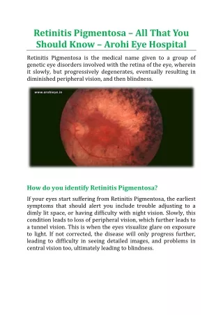
Retinitis pigmentosa (RP) is a group of inherited diseases that damage the light-sensitive rods and cones located in the retina, the back part of our eyes. Rods, which provide side (peripheral) and night vision are affected more than the cones that provide color and clear central vision.
Signs of RP usually appear during childhood or adolescence. The first sign is often night blindness followed by a slow loss of side vision. Over the years, the disease will cause further loss of side vision. As the disease develops, people with RP may often bump into chairs and other objects as side vision worsens and they only see in one direction - straight ahead. They see as if they are in a tunnel (thus the term tunnel vision).
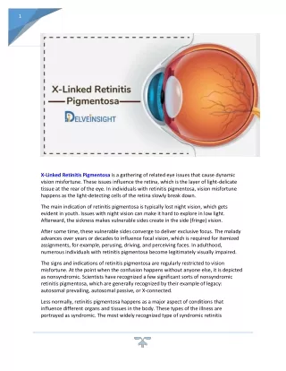
RP is an inherited disorder that results from harmful changes in any one of more than 50 genes. These genes carry the instructions for making proteins that are needed in cells within the retina, called photoreceptors.Fortunately, most cases of retinitis pigmentosa take a long time to develop and vision loss is gradual. It may take many years for loss of vision to be severe.The retina is the light-sensitive tissue at the back of the eye that contains photoreceptors and other cell types
How is RP inherited? It is important to know a little more about genes and how they are passed from parent to child. Genes are bundled together on structures called chromosomes. Each cell in our body contains 23 pairs of chromosomes. One copy of each chromosome is passed by a parent at conception through egg and sperm cells. The X and Y chromosomes, known as sex chromosomes, determine whether a person is born female (XX) or male (XY). The 22 other paired chromosomes, called autosomes, contain the vast majority of genes that determine non-sex traits. RP can be inherited in one of three ways:
Autosomal recessiveInheritance • Autosomal dominant Inheritance • X-linked Inheritance • The symptoms of RP typically appear in childhood. They often have difficulty in getting around in the dark. It can also take abnormally long periods of time to adjust to changes in lighting. As their visual field becomes restricted, patients often trip over things and appear clumsy. People with RP often find bright lights uncomfortable, a condition known as photophobia..
It is caused due to many gene mutations that cause the disorder, its progression can differ greatly from person to person. Some people retain central vision and a restricted visual field into their 50s, while others experience significant vision loss in early adulthood. Eventually, most individuals with RP will lose most of their sight
Diagnosis of Retinitis Pigmentosa: Eye care professional will use an ophthalmoscope, a tool that allows for a wider, clear view of the retina. This typically reveals abnormal, dark pigment deposits that streak the retina. Other tests for RP include: • Electroretinogram (ERG) • Visual field testing. • Genetic testing.
Electroretinogram (ERG). It measures the electrical activity of photoreceptor cells. This test uses gold foil or a contact lens with electrodes attached. A flash of light is sent to the retina and the electrodes measure rod and cone cell responses. People with RP have a decreased electrical activity, reflecting the declining function of photoreceptors. Visual field testing: To determine the extent of vision loss, a clinician will give a visual field test. The person watches as a dot of light moves around the half-circle (180 degrees) of space directly in front of the head and to either side. The patient pushes a button to indicate that he or she can see the light. This process results in a map of their visual field and their central vision.
Genetic testing. In some cases, a clinician takes a DNA sample from the person to give a genetic diagnosis. It is available for a limited number of patients with RP through NEI (National Ophthalmic Disorder Genotyping and Phenotyping Network) (eye GENE).
Treatment Currently, there is no cure for RP, but there is research that indicates that vitamin A and lutein may slow the rate at which the disease progresses. Your doctor of optometry can give you more specific information on nutritional supplements that may help you. Also, there are many new low vision aids, including telescopic and magnifying lenses, night vision scopes as well as other adaptive devices, that are available that help people maximize the vision that they have remaining. An optometrist, experienced in low vision rehabilitation, can provide these devices as well as advice about other training and assistance to help people remain independent and productive. Because it is an inherited disease, research into genetics may one day provide a prevention or cure for those who have RP.
Classroom and Teachers Provide a copy if notes as student may have difficulty coping from the board. Well lit area of room, however be aware of glare. Allow additional time to complete assignments or shorten them Using framing technique to help student focus on one problem at a time Seat student in middle or back of room to help them increase their visual field. Student : Move to find best vision field. Use low vision devices if necessary. Infared blocking sunglasses.
Parent/Family • Get counseling to help understand this eye condition. • Be support of and have other family members check. • Ask questions.