Understanding the Maternal Bony Pelvis Anatomy
700 likes | 989 Views
Learn about the structure, divisions, and important measurements of the maternal bony pelvis in obstetrics and gynecology.

Understanding the Maternal Bony Pelvis Anatomy
E N D
Presentation Transcript
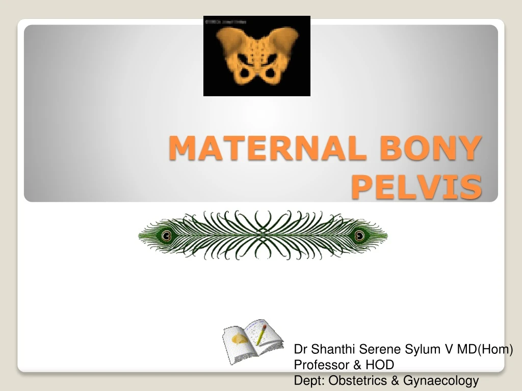
MATERNAL BONY PELVIS Dr Shanthi Serene Sylum V MD(Hom) Professor & HOD Dept: Obstetrics & Gynaecology
Bones are – • Iliac bones – 2 • Sacrum - 1 • Coccyx - 1 Joints are – • Sacro iliac joints – 2 • Sacro coccegial jt – 1 • Symphysis pubis - 1 An articulated pelvis consist of 4 bones & they are united by 4 joints.
False pelvis Or the greater pelvis or pelvis major True pelvis Or The lesser pelvis Or Pelvis minor Anatomically pelvis is divided into 2
1. upper border of symphysis pubis • 2. Pubic crest • 3. Pubic tubercle • 4. Pectineal line • 5. Iliopubic eminence • 6. Iliopectineal line • 7. Sacroiliac articulation • 8. Anterior border of the ala of sacrum • 9. Sacral promontory Bony land marks of pelvic brim from anterior to posterior -
It is formed by the iliac portion of the innominate bone. • It got only a little obstetric significance. • Practically it’s measurements are not taken now. • It’s function is to support uterus during pregnancy. • Boundary – Posteriorly – Lumbar vertibrae Laterally - iliac fossa anteriorly - Anterior abdominal wall False pelvis
1. interspinous diameter • 2. Intercristal diameter • 3. Baudeloque’s diameter / Ext conjugate • 4. Inter trochanteric diameter • (These diameters have no clinical values) Measurements of False pelvis
Transverse distance between outer lips of anterior superior iliac spines. • about 22 - 25cm • ie (9 -10”). 1. Interspinous diameter
Transverse distance between outer lips of iliac crests at the widest part . • about 25 – 28 cm. • ie 10 – 11” 2. Intercristal diameter
Antero posterior distance between tip of last lumbar vertebra & mid point of superior border of symphysis pubis. • It measures 19 cm.( 71/2”) • Baudeloque believed that true conjugate can be obtained by deduction of 3 ¼” from External conjugate 3. Baudeloque’s diameter or External conjugate
Maximum width between greater trochanters. • about 31cm. 4. Inter-trochanteric diameter
This forms the bony birth passage of foetus. • It is shallow in front & deep posteriorly. • Bounded anteriorly by symphysis pubis which measures 4 cm posteriorly by sacrum & coccyx measuring 11.5 cm True pelvis
1. Pelvic inlet / brim 2. Pelvic cavity 3. Pelvic outlet For descriptive purpose it is devided into 3
Inlet is formed by – • 1. upper border of symphysis pubis • 2. Pubic crest • 3. Pubic tubercle • 4. Pectineal line • 5. Iliopubic eminence • 6. Iliopectineal line • 7. Sacroiliac articulation • 8. Anterior border of the ala of sacrum • 9. Sacral promontory 1. Pelvic inlet / brim
Shape • Plane • Inclination • Sacral angle • Axis • Diameters Things to be known in thePelvic inlet / brim
heart-shaped or rounded with slight antero posterior flatening due to forward projection of sacral promontory. Shape -
It is the space of pelvic brim through which an imaginary plane is drawn. Plane or superior strait
Pelvis is tilted forward in the erect posture. • So the plane of inlet makes an angle of 55 with the horizontal plane. This angle is called angle of inclination. • angle of inclination can be measured in another way too. • Ie. the angle formed between the plane of inlet & the front of the body of V lumbar vertebra which measures 135 . 55 Inclination 55 It can be measured radiographically.
If the angle of inclination is increased due to sacralization of 5th lumbar vertebra, it is called high inclination. • It got obstetric significance like – It produce delay in engagement because the uterine axis fails to coincide with the inlet. It favours occipito posterior position. It produce difficulty in descent of the head due to long birth canal & flat sacrum. High inclination
If the angle of inclination is lessened due to lumbarizationof 1st piece of sacral vertebra, then it is called low inclination. • It facilitates early engagement. low inclination
It is the angle formed by plane of brim & 1sttwo pieces of sacrum. • Normaiiy it is more than 90 . • Narrow sacral angle suggest flat sacrum in funnel pelvis. Sacral angle
Mid perpendicular line drawn to the plane of inlet directed downward & backwards. • When extended, this line passes through the umbilicus to coccyx. • This Axis of inlet should coincide with the uterine axis for the easy passage of foetus through the brim. Axis
1. Antero posterior diameter 2. Obstetric conjugate 3. Diagonal conjugate 4. Oblique diameter 5. Transverse diameter Diameters
Extends from midpoint of sacral promontory to midpoint of upper border of symphysis pubis. • Measures 11 cm (4 ¼”) • It cannot be estimated directly. • It is indirectly measured by deducting 1.2 cm (1/2”) from the diagonal conjugate. • Syn – True conjugate, anatomical conjugate, conjugate vera Antero diameter posterior
It is the distence between midpoint of sacral promontory to prominent bony projection in the mid line on the inner surface of the symphysis pubis. • It is the shortest Antero posterior diameter of the inlet. • It Measures 10 cm (4”) • It cannot be clinicaly estimated . • It is indirectly measured by deducting 1.5 - 2 cm (3/4”) from the diagonal conjugate.. 2. Obstetric conjugate
It is the distence between the lower border of symphysis pubis to the midpoint on sacral promontory. • It Measures 12 cm (4 3/4”) • It is measured during pelvic assessment in late pregnancy or in labour. 3. Diagonal conjugate
Place pt in dorsal position. • Take aseptic precautions. • Extended index & middle fingers of gloved rt hand are introduced into vagina. • Try to palpate through posterior vaginal fornix against hollow of the sacrum from below upwards with an attempt to reach the sacral promontory through the sacral concave curve. { it is difficult in normal pelvis} • The point at witch the bone recedes from the fingers is the sacral promontory. • Mark a point just below the symphysis pubis over the rt gloved finger. • The distence between the sacral promontory & the Marked point gives the Diagonal conjugate . How to measure Diagonal conjugate ?
It measures from one sacroiliac joint to opposite iliopectenial eminance. • They are two in no. • Rt o d – Rt to Lt. • Lt o d - Lt to Rt • Measures 12 cm • (4 ¾”) 4. Oblique diameter L O R O
Maximum distance between two farthest points on the iliopectenial eminance. • It measures 13 cm • (5 ¼”) • It lies closer to sacral promontory at a distance of 4 cm 4 cm 5. Transverse diameter
It is the distence between the mid point of the sacral promontory to iliopectenial eminance. • It represcents the space occupied by the biparietal diameter of the head while negotiating the brim in flat pelvis. 6. Sacro cotyloid diameter
plane of greatest pelvic dimension Shape boundary Diameters Things to be known in the Cavity are -
Cavity is the segment of pelvis bounded above by the inlet & below by plane of least pelvic dimensions. • Shape is almost round. Cavity
it is the plane of greatest pelvic dimension & it is the most roomy plane in true pelvis. • Planeextend from Ant - midpoint of posterior surface of symphysis pubis Post - junction of 2nd & 3rd sacral vertebrae Later – ischial bone over middle of acetabulum. • Shape of plane – Round • Diameters Anteroposterior – 4 ¾”(12 cm) Oblique – 4 ¾”(12 cm) Transverse – 4 ¾”(12 cm) Plane
Plane of least dimensions. Extends throughlower margine of symphysis pubis,tip of the sacrum and ischial spines.Antero-post diameter about 11.5cm, Transverse diamete about10.5cm. Plane
The lower circumference of the pelvis is very irregular; the space enclosed by it is named the inferior aperture or outlet • And is bounded behind by the point of the coccyx, and laterally by the ischial tuberosities. • These eminences are separated by three notches: one in front, • The pubic arch, formed by the convergence of the inferior rami of the ischium and pubis on either side.
The other notches, one on either side, are formed by the sacrum and coccyx behind, the ischium in front, and the ilium above; they are called the sciatic notches; • In the natural state they are converted into foramina by the sacrotuberous and sacrospinous ligaments. • When the ligaments are in situ, the inferior aperture of the pelvis is lozenge-shaped, bounded, in front, by the pubic arcuate ligament and the inferior rami of the pubes and ischia; laterally, by the ischial tuberosities; and behind, by the sacrotuberous ligaments and the tip of the coccyx.
Obstetrical Outlet Boundary Shape Plane Axis Diameters Antero posterior Transverse Posterior sagittal Anatomical Outlet Boundary Shape Plane Axis Diameters Antero posterior Transverse Posterior sagittal Subpubic angle Pubic arch Outlet
It is the segment of pelvis bounded by the plane of the least pelvic dimensions & below by the anatomical outlet. • Its anterior wall is deficient at the pubic arch. • Lateral walls are formed by ischial bones. • Posterior wall forms coccyx • Orbony outlet. • bounded Anteriorly - inferior border of symphysis pubis Laterally – ischiopubic rami , ischial tuberosity, , sacrotuberous and sacrospinous ligaments posteriorly - tip of coccyx. It consist of two triangular planes with a common base formed by joining 2 ischial tuberosities. Ant apex - at inferior border of pubic arch. Post apex – at the tip of the coccyx. Obstetrical OutletAnatomical Outlet
Boundary Anteriorly - inferior border of symphysis pubis, Laterally - pubic bone, ischium , sacrotuberous and sacrospinous ligaments posteriorly - tip of coccyx. Planes of pelvic outlet They lie on two triangular planes with base joining two ischial tuberosites due to downward projection of the ischial tuberosites. Shape - oval PELVIC OUTLET
Antero posterior - • 5 ¼” (13cm) • extends from mid point of inferior border of symphysis pubis to tip of coccyx. • posterior point becomes tip of sacrum when coccyx is displaced backward during delivery of fetal head. • OBLIQUE - 4 ¾” (12cm) Diameters
Transverse - measures 4¼” (10.6cm) measuring from medial border of the ischail tuberosities.This is called TRANSVERSE DIAMETER OF OUTLET (TDO) • It can be clinically measured.
Post sagital diameter Distance from mid point of line between ischial tuberosities and external surface of tip of scrum about 7.0cm.
It is formed by the descending rami of pubic bones. • It measures 90 degree or more. • subpubic arch is wide and round disc of 9.4cm diameters. • Diameter of well flexed vertex can pass through pubic arch at a distance of 1cm from midpoint of inferior border of symphysis. • This distance is called waste space of Morris. 9.4cm Sub pubic angle
If the waste space Morris is more than 1cm due to narrow subpubic angle, available antero posterior diameter of outlet becomes less, and fetal head had to pass injuring perineum or even gets arrested .
MIDPELVIS is the narrowest segment of true pelvis lying between obstetric outlet below and roomy pelvic cavity above. MIDPELVIS
Plane. Plane at midpelvis is called plane of least pelvic dimension. • Boundaries- anteriorly lower border of symphysis pubis, inner aspect of inferior ramus and obturator foramen, ischialspine, sacrospinous ligament and tip of sacrum. • Ischail spines form two important landmarks of this plane. • Shape of plane-anteroposterior oval. MIDPELVIS