Download

1 / 26
280 likes | 377 Views
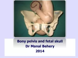
THE BONY PELVIS. DR. AHMED ABDULWAHAB Assistant Professor, Consultant OBGYN Department. In women the pelvis has special form that adapts to childbearing . It is composed of four bones . The sacrum coccyx and two innominate bones ..

E N D
THE BONY PELVIS DR. AHMED ABDULWAHAB Assistant Professor, Consultant OBGYN Department
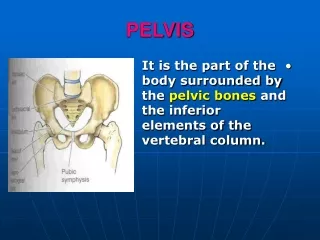
In women the pelvis has special form that adapts to childbearing . • It is composed of four bones . • The sacrum coccyx and two innominate bones .. • The innominate bone is formed by the fusion of the ilium ,ischium, and pubis
The true pelvis is the portion important in childbearing , is bounded above by promontory and alae of the sacrum the lineaterminalis and the upper margin of the pubic bone , and below by the pelvic outlet . • Ischial spines are of great obstetrical importance because it is the shortest pelvic diameter and has a valuable landmarks in assessing the level of the presenting part of the fetus
The sacrum form the posterior wall of the pelvis and it is curved to accommodate the rotating head . • The promontory may be felt on vaginal examination and provide a landmark for clinical pelvimetry
Pelvic joints • Symphysis pubis where pelvic bones are joined together anteriorly • Sacroiliac joint where pelvic bones are joined posteriorly .
Planes and diameters of the pelvis • Four imaginary planes • 1- the plane of pelvic inlet • 2-the plane of pelvic outlet • 3- the plane of mid pelvis • 4 –the plane of greatest pelvic dimensions
Pelvic shapes • 1-gynecoid pelvis the inlet is slightly oval or rounded , the ischial spines are not prominent , the pubic arch is wide , sacrum is well curved . • Android pelvis is a deep and convergent with prominent ischial spines , narrow sub-pubic arch and straight sacrum .
Pelvic inlet measurement • Diagonal conjugate it is the distant from the sacral promontory to the lower margin of the symphysis pubis. • True conjugate from sacral promontory to upper border of symphysis pubis • Obstetric conjugate from sacral promontory to mid of posterior aspect of symphysis pubis subtract 1.5-2.0 cm from diagonal conjugate
The mid pelvis at the level of ischial spines the inter- spinous diameter is 10 cm . • Pelvic outlet clinically it is the distant between the ischialtuberosities it is around 8.0 cm
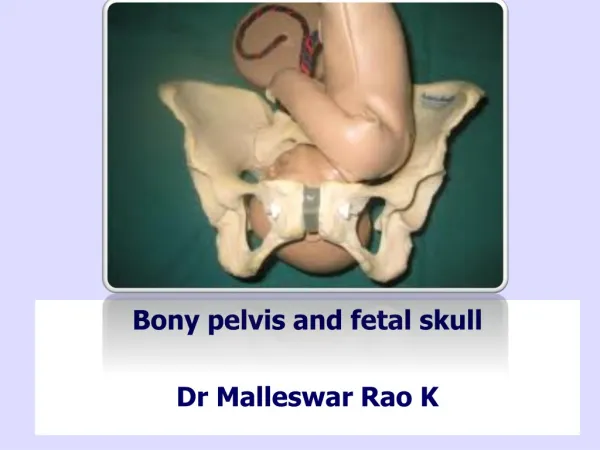
THE FETAL SKULL • BONES • Two frontal bones separated by frontal suture. • Two parietal bones separated by sagittal suture . • Two coronal sutures between frontal and parietal bones . • Two lambdoid sutures between parietal and occipital bone .
Sutures meet at an irregular space forms which is enclosed by a membrane called fontanel . • Anterior fontanel is a lozenge shape between the two frontal and two parietal bones usually it is opened . • Posterior fontanel at the junction of the two parietal bones and occipital bone . • It gives an important information concerning presentation and position of the fetus.
Fetal head diameters • Subocipoto-bregmatic 9.5 cm vertex presentation. • Submento-bregmatic 9.5 cm face presentation. • Mento-vertical 12.5 brow presentation . • Biparietal diameter 9.5cm . • Occipto-frontal 10.5 cm
Occipital bone is the landmark in vertex presentation. • Mentum is landmark for face presentation, • Frontal bone is land mark for brow presentation
labour Definition. It is the onset of painful, regular ,contractions, more than one every ten minutes. With progressive cervical effacement and dilatation accompanied by descend of the fetal presenting part.
Stages of labor Labor is divided in to three stages. 1st stage from diagnosis of labor till full dilatation of the cervix. 2nd stage of labor from full dilatation of the cervix till delivery of the fetus. 3rd stage from delivery of the fetus until delivery of the placenta.
The duration of labor Primigravida about 12 hours . Multigravida 8.0 hours The moral of most women deteriorate if labor is prolonged . There is greater incidence of fetal hypoxia after long labor. Greater incidence of operative vaginal delivery.
Mechanisim of labor It is a series of changes in position and attitude that the fetus undergoes during its passage through the birth canal. ENGAGEMENT. It is when the widest diameter of the head has passed successfully through the inlet that is when the bi-parietal diameter passed to the level of the ischial spines
DESCENT. It is secondary to uterine action in 1st and early phase of 2nd stage of labor . FLEXION When the head descent to the narrow mid-cavity flexion should occur.
INTERNAL ROTATION . The shape of the bony pelvis and direction of the pelvic floor muscles in addition to the well flexed head will help the head to rotate the head into the occipito anterior position . In a well flexed head the occiput will meet the pelvic floor and will guide the direction of the rotation
EXTENSION. The head is deliver by extension first the bregma ,face , and chin appear in succession over the posterior vaginal opening and perineal body. RESTITUTION. As soon as the head escape from the vulva the head aligns itself with the shoulder
EXTERNAL ROTATION. In order to deliver the shoulders have to rotate into the direct anterior- posterior plane . The doctor will rotate the head making the face of the fetus looking to medial aspect of the maternal thigh .
Delivery of the shoulders . The anterior shoulder is under the symphysis pubis and deliver first ,and the posterior shoulder deliver subsequently
THIRD STAGE OF LABOR . Separation of the placenta occurs because of the reduction of the volume of the uterus due to the uterine contraction and retraction