Download
1 / 8
80 likes | 134 Views
Explore the intricate structure of the auditory system, including the cochlea, vestibule, and SCCs within the bony labyrinth. Discover the roles of various components such as the modiolus, osseus spiral lamina, and tectorial membrane in auditory perception. Gain insights into the inner and outer hair cells, their functions in transducing mechanical movement into electrical signals, and the auditory nerve pathway. A mnemonic guide (ECOLI) simplifies the pathway from stimulation to auditory cortex response. Unravel the complexity of auditory regions within the transverse gyri of Heschl.

E N D
Sameer Ahmed 9/25/2013 Cummings Ch 128: Auditory System Anatomy
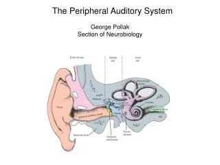
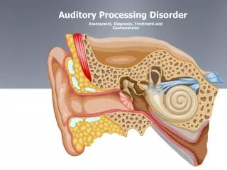
Bony labyrinth • Hardest bone of the body • Comprised of the cochlea, vestibule, and SCC's • Cochlea • 2 and ¾ turns (also will see 2 and ½ turns in other texts) • Modiolous • Core of the cochlea • Highly porous bone to allow passage of auditory nerve fibers to hair cells • Osseus spiral lamina • Extends from the modiolous • Divides upper and lower cochlear chambers into scala vestibuli and scala tympani • Also serves as a point of attachment for the basilar membrane (separates scala media from scala tympani)
Membranous labyrinth – houses organ of corti • Superiorly by Reissner's membrance, Inferiorly by basilar membrane, laterally by spiral ligament • Stria Vascularis: highly vascular tissue responsible for metabolic environment of scala media
Tectorial membrane • Lies over the inner and outer hair cells • Gelatinous structure • Deiter's cells, Hansen cells, Claudius' cells → supporting cells of the Organ of Corti • Perilymph: High Na, Low K • Between osseus and membranous labyrinth • Endolymph: High K, Low Na • Found within the membranous labyrinth • Enlarged vestibular aqueduct, defined as a diameter larger than 1.5 mm at the vestibular aqueduct midpoint • Minor head trauma can lead to progressive SNHL
The inner and outer hair cells function as receptor cells that transduce mechanical movement into an electrochemical signal to stimulate the auditory nerve. • 3500 inner hair cells • 12,000 outer hair cells • Afferent innervation of these hair cells comes from 30,000 auditory nerve fibers (cochlear portion of CN 8) • Type I fibers are bipolar, large diameter, and myelinated, • Constitute nearly 95% of the total number of fibers. • Each type I fiber has a direct and independent synapse on the body of a single inner hair cell, and each inner hair cell is innervated by approximately 20 such fibers. • Type II fibers constitute the remaining 5%, and are smaller and may be myelinated or unmyelinated. • Type II fibers synapse directly on the outer hair cells, with a single fiber diverging to form branches that synapse with multiple other outer hair cells.
Auditory Nerve Pathway Mnemonic • ECOLI • E ighth nerve • C ochlear nucleus • superior O livary nucleus • L ateral lemniscus • I nferior colliculus
Pathway 1) Stimulation of hair cells from vibration of basilar membrane stimulates bipolar neurons of spiral ganglion that form cochlear division of CN 8 2) Cochlear neurons then synpase in the cochlear nuclei (anterior ventral, posterior vental, and dorsal) 3) Bilateral innervation from the cochlear nucleus (via acoustic striae) synpase on the superior Olivary nucleus 4) Superior olivary nuclei → lateral lemniscus → inferior colliculus → thalamus (medial geniculate body) –> auditory cortex at Sylvian fissure of temporal lobe (Brodmann area 41) • Auditory regions within the transverse gyri of Heschl