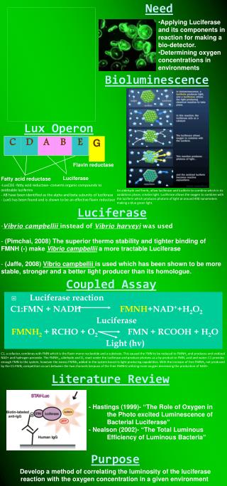
Luciferase
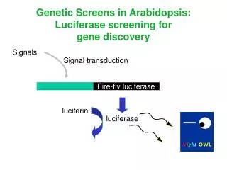
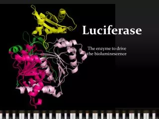
Luciferase. The enzyme to drive the bioluminescence. Introduction to Luciferase. Luciferase, also be called as luminase, is a generic terms for enzymes that catalyze the process of bioluminescence in nature. Luciferin + ATP Oxyluciferin + AMP + Light

Luciferase
E N D
Presentation Transcript
Luciferase The enzyme to drive the bioluminescence
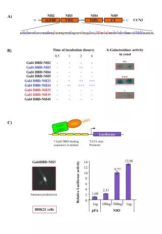
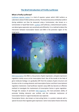
Introduction to Luciferase • Luciferase, also be called as luminase, is a generic terms for enzymes that catalyze the process of bioluminescence in nature. • Luciferin + ATP Oxyluciferin + AMP + Light • The research of luciferase began at the fifth ties in twentieth century. The first luciferase to be cloned and also the first to be structurally characterized is bacterial luciferase. Luciferase
Classification • Bacterial Luciferase: • http://filebox.vt.edu/users/chagedor/biol_4684/Methods/bacteria.gif • Firefly Luciferase: • http://www.mrnussbaum.com/firefly.gif • Other organism: • http://www.flmnh.ufl.edu/fish/education/adapt/biolum.JPG
Catalytic mechanism of Luciferase Step 1: Luc + LH2 + ATP Mg2+ Luc LH2 – AMP + PPi Luc Luc LH2 – AMP Luc Step 2: Luc LH2 – AMP + O2 Luc Oxyluciferin (EES) + AMP + CO2 O2 Step 3: Luc Oxyluciferin (EES) Luc Oxyluciferin + green light
Fig.10. The binding of AMP with luciferase (PDB ID: 2d1q) generated with PyMOL. The view shows AMP binding with luciferase [Nature 440: 372-376] Fig.11. (left) The binding of AMP with luciferase (PDB ID: 2d1q) generated with PyMOL. The Gly341, Gly 343, Tyr 342 and Ala319 of Firefly Luciferase work together to form a pocket to hold the adenine ring of AMP [Nature 440: 372-376] Step 1: the formation of Luc LH2 – AMP 1. The formation of Luc-AMP complex:
Fig.12. The Luc LH2 – AMP active intermediate (PDB ID: 2d1s) generated with PyMOL. The picture shows the Arg220, Phe249, Ser349 and Ala350 patch like a pocket to hold the Luciferin to Firefly luciferase to form the intermediate, also is shown in this picture is the binding of Lys531 to reduce the energy of the transition state. [Nature 440: 372-376] Fig.13. (left) The Luc LH2 – AMP active intermediate (PDB ID: 2d1s) generated with PyMOL. The picture shows the binding of AMP and Luciferin to Firefly luciferase to form the intermediate [Nature 440: 372-376] Step 1. The formation of Luc LH2 – AMP 2. The formation of Luc LH2 – AMP active intermediate
Fig.13. Intermediate change to Oxyluciferin and AMP (PDB ID: 2d1r) generated with PyMOL. The picture shows how the Luc LH2 – AMP intermediate change to Oxyluciferin and AMP. [Nature 440: 372-376] Fig.13. The OxyLuc and AMP binding to Luciferase (PDB ID: 2d1r) generated with PyMOL. The picture shows the binding of AMP and OxyLuciferin to Firefly luciferase. [Nature 440: 372-376] Step 2: The formation of Luc Oxyluciferin (EES) and the leaving of AMP
Referrence • 1. Toru Nakatsu, Susumu Ichiyama, Jun Hiratake, Adrian Saldanha, Nobuyuki Kobashi, Kanzo Sakata and Hiroaki Kato. Structural basis for the spectral difference in luciferase bioluminescence. Nature 440, 372-376