Download
1 / 2
20 likes | 233 Views
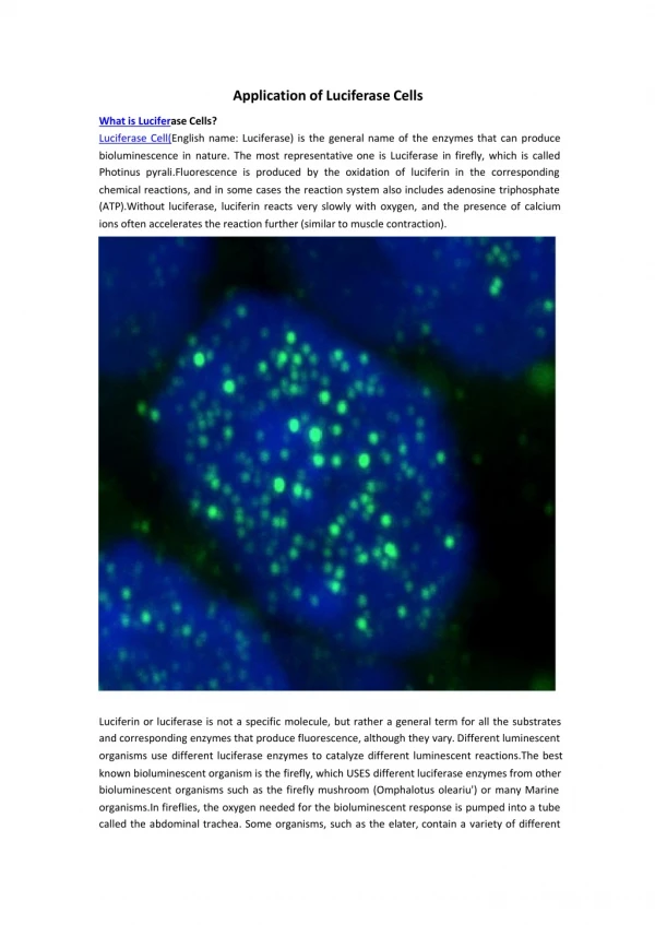
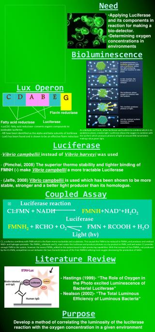
Need. G. C. D. A. B. E. Luciferase. Fatty acid reductase. Flavin reductase. Applying Luciferase and its components in reaction for making a bio-detector. Determining oxygen concentrations in environments . Bioluminescence. Lux Operon.

E N D
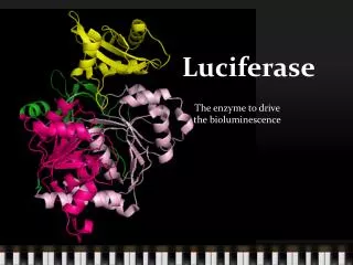
Need G C D A B E Luciferase Fatty acid reductase Flavin reductase • Applying Luciferase and its components in reaction for making a bio-detector. • Determining oxygen concentrations in environments Bioluminescence Lux Operon -LuxCDE -fatty acid reductase- converts organic compounds to oxidizable luciferins - AB have been identified as the alpha and beta subunits of luciferase - LuxG has been found and is shown to be an effective flavin reductase An aldehyde and fmnh2 allow luciferase and luciferin to combine which in its oxidations phase, creates light. Luciferase allows the oxygen to combine with the luciferin which produces photons of light at around 490 nanometers making a blue green light. Luciferase • Vibrio campbellii instead of Vibrio harveyi was used • (Pimchai, 2008) The superior thermo stability and tighter binding of FMNH (-) make Vibrio campbellii a more tractable Luciferase • (Jaffe, 2008) Vibrio campbellii is used which has been shown to be more stable, stronger and a better light producer than its homologue. Coupled Assay • Luciferase reaction C1:FMN + NADH FMNH+NAD++H2O2 Luciferase FMNH2+ RCHO + O2 FMN + RCOOH + H2O Light (hv) C1, a cofactor, combines with FMN which is the flavin mono nucleotide and a substrate. This caused the FMN to be reduced to FMNH2 and produces and oxidized NAD+ and hydrogen peroxide. The FMNH2, aldehyde and O2 react under the luciferase and produce photons as a by product to FMN, acid and water. C1 provides enough FMN to the system, however the excess FMNh2 added to the system boost its light producing capabilities. With the increase of free FMNH2 not produced by the C1:FMN, competition occurs between the two channels because of the Free FMNH2 utilizing more oxygen decreasing the production of NAD+. Literature Review - Hastings (1999)- “The Role of Oxygen in the Photo excited Luminescence of Bacterial Luciferase” - Nealson (2002)- “The Total Luminous Efficiency of Luminous Bacteria” Purpose Develop a method of correlating the luminosity of the luciferase reaction with the oxygen concentration in a given environment
Results • Standard curves were calculated based on the results of the various test • Equations of the curves calculated by breaking graphs in to parts and creating trend lines of either polynomial equations or algorithmic equations in order to define graphs • Equations shown are for trials containing no extra FMN added to the system along with HPA and 9.2 micro molar solution of C1 and Lux y = -0.000x4 + 0.008x3 - 0.141x2 + 1.356x + 20.69R² = 1 Equation of curve with range less than 146 a.u y = 0.001x3 - 0.218x2 + 16.38x - 246.4R² = 1 Equation of curve with range greater than 146a.u Discussion • Adding free FMN causes the increase of hydrogen peroxide which decreases the light production path. • Taking away free FMN created a higher yield of light since the wasteful FMNH- consumption to produce H2O2 was lessen. • FMNH2 in higher oxygen concentrations reacts with oxygen rather than aldehyde to produce H2O2 which inhibits FMNH2 from going into the luciferase reaction Conclusion • The oxygen concentration of the environment has an impact on the luminosity of Vibro campbellii luciferase • The luminosity of the reaction can be used to correlate the oxygen concentration of a given environment • 0 Future Studies • Using increasing amounts of concentrations of Lux in order to obtain a more defferiantable curve • Using different strains and species of Lux in order to test for their effect on the system Branchini, BR; Magyar, RA; Murtiashaw, MH; Anderson, SM; and others. (1999) Site-directed mutagenesis of firefly luciferase active site amino acids: A proposed model for bioluminescence color. Biochemistry 38:13223-13230. Chaiyen, Pimchai. LuxG is a functioning flavin reductase for bacterial luminescence. Department of Biochemistry and Center for excellence in Protein structure and function. 9/15/07. Dave and Barie Deo, S.K., Mirasoli, M. and Daunert, S. (2005) Bioluminescence resonance energy transfer from aequorin to a fluorophore: an artificial jellyfish for applications in multianalyte detection. Analytical and Bioanalytical Chemistry, 381: 1387-1394. Hastings, Woodland. Chemistries and colors of bioluminescent reaction: a review. Department of Molecular and Cellular Biology, Harvard University. 1005. Elsevier Science V.V . SSDI 0378-1119 Harvey, Newton. The Total Luminous Efficiency of Luminous Bacteria. The journal of General Physiology. September . 1975 Hastings. Woodland. The Role of Oxygen in the Photo excited Luminescence of Bacterial Luciferase. Department of Molecular and Cellular Biology. Harvard. The journal of Biological Chemistry. Vol 242. NO 4 Issue February. Pg 720-726 Jaffe, Lionel. Bioluminescence. May 2008. http://www.lifesci.ucsb.edu/~biolum/ Jablonski, Edward. Immobilization of bacterial luciferase and FMN reductase on glass rods. Pro. Natl. Acad. Sci USA. Vol. 73, no 11 pp 3848-3851. November 1976 McElroy, W. D., and M. DeLuca. 1985. Firefly luminescence, p.387-399. In J. G. Burr (ed.), Chemi- and bioluminescence.Marcel Dekker, Inc., New York. Nakamura, Makiko. Construction of streptavidin luciferase fusion protein for ATP sensing with fixed form. Biotechnology Letters 26: 1061-1066. 2004. Pg 1061-1072 Ron, Eliora. Biosensing enivornmental pollution. Current Opinion in Biotechnology, Volume 18. Issue 3, June 2007. Pg 252-256 Shimomura, O. (2005) The discovery of aequorin and green fluorescent protein. Journal of Microscopy, 217: 3-15. Tannous, B.A., Kim, D.-E., Fernandez, J.L., Weissleder, R. and Breakefield, X.O. (2005) Codon-Optimized Gaussia Luciferase cDNA for Mammalian Gene Expression in Culture and in Vivo. Molecular Therapy, 11: 435-443. Taylor, Amanda. Bioluminescence detection of ATP release mechanisms in epithelia. American Physiological Society. 1998. Pg. c1391. Ajpcell.physiology.org Wet, Jeffery. Firefly Luciferase Gene: Structure and Expression in Mammalian Cells. 1986. Departments of Biology' and Chemistry,2 University of California. MOLECULAR AND CELLULAR BIOLOGY Verhaegen M. and Christopoulos, T.K. (2002) Recombinant Gaussia luciferase. Overexpression, purification and analytical application of a bioluminescent reporter for DNA hybridization. Analytical Chemistry, 74: 4378-4385 http://www.pubmedcentral.nih.gov/articlerender.fcgi?artid=1932783 Bibliography