Download

1 / 23
230 likes | 366 Views
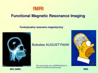
This article explores the neurobiological underpinnings of reading through advanced fMRI techniques. Focusing on different slice orientations such as sagittal, coronal, and axial, it assesses brain areas involved in semantic processing during auditory and visual sentence tasks. Specific regions, including the temporoparietal and occipitotemporal areas, are analyzed for their roles in mapping orthographic, phonological, and semantic representations, as highlighted in studies by Pugh et al. (2000) and others. The impact of functional connectivity on reading abilities among various demographic groups is also discussed.

E N D
slice orientations sagittal coronal axial
SPGR Anatomics (1) resolution in-plane 0.9375 mm2 through-plane 1.5 mm
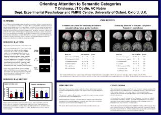
Auditory versus Visual Sentence Task common print(red) speech(blue) R - L Constable, Pugh et al., (NeuroImage 2004)
An Initial Neurobiological Reading Model (Pugh et al. 2000) • TEMPOROPARIETAL • (DORSAL) • Areas: • supramarginal, angular, superior temporal (Wernicke’s) gyri • Hypothesized Function: • Mapping of orthographic to phonological and semantic representations • ANTERIOR • Areas: • inferior frontal gyrus (including Broca’s area) • One Hypothesized Function: • Articulatory recoding OCCIPITOTEMPORAL (VENTRAL) Areas: occipitotemporal juncture, middle and inferior temporal gyri Hypothesized Function: Linguistically structured memory- based fast word identification system (posterior aspect = “word-form” area)
Auditory versus Visual Sentence Task common print(red) speech(blue) temporoparietal anterior occipitotemporal R - L Constable, Pugh et al., (NeuroImage 2004)
Vasculature Menon & Kim, Trends in Cognitive Sciences, 3(6), 1999.
Vasculature Menon & Kim, Trends in Cognitive Sciences, 3(6), 1999.
Simulated Hemodynamic Response (1) mean = 5000 sd = 100 effect size 0-1%, 0-50 points Gamma function tau=1.08; n=3; delay=3
Variability of the HRF (1) Meizin et al., 2000
Simulated Hemodynamic Response (2) Noise SD = 0 Noise SD = 10 Noise SD = 100
Estimating the Response (3):Simple Subtraction with Overlap Dale & Buckner, 1997
Linear Deconvolution Neuronal activity is modeled as a sequence of point events. slide courtesy of Matthew Brown, University of Western Ontario
Functional Connectivity activations in Shaywitz et al. 2002 older good readers 74 good readers 7-18 yrs 70 dyslexic readers 7-18 yrs
Functional Connectivity seed voxel correlations older good readers
Functional Connectivity Younger Non-Impaired Older Non-Impaired univariate correlations Older Dyslexics Younger Dyslexics