Download

1 / 27
270 likes | 469 Views
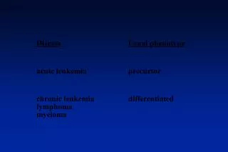
LEUKEMIA—HEMATOLOGY {S1}. BY RANJEET RAMAN. Leukemia is divided into acute vs. chronic, and myeloid vs. lymphoid. The transforming event in leukemia affects a progenitor cell.

E N D
LEUKEMIA—HEMATOLOGY {S1} BY RANJEET RAMAN
Leukemia is divided into acute vs. chronic, and myeloid vs. lymphoid. The transforming event in leukemia affects a progenitor cell. • Chronic leukemias generally consist of proliferation of mature elements that resemble normal blood and marrow elements. • Acute leukemias generally consist of proliferation of blasts, with an arrest of normal maturation.
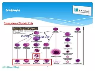
Chronic Myeloid Leukemia (CML) • CML is characterized by Granulocytosis in the peripheral blood. White blood counts are often between 50,000 and 200,000. Mature and immature myeloid cells, including bands, metamyelocyte, and promyelocyte, are seen peripherally.
Although the first cancer cell was a stem cell, mature cells dominate in CML. • The bone marrow is extremely hypercellular with heterogeneous myeloid elements. • Megakaryocytes are increased as well, but are smaller than normal.
Patients may be asymptomatic, but are shown to have huge WBC counts and splenomegaly. • Other tissues may also be infiltrated with myeloid cells. This is called “chloroma.”
**The most important lab finding is t(9;22) translocation, which is called the Philadelphia chromosome. This creates the bcr-abl oncogene. • Gleevec directly and specifically inhibits the bcr-abl tyrosine kinase, practically curing CML.
CML often starts chronically but then accelerates into a blast crisis characterized by more immature myeloid cells. It can be thought of as a myelo-accumulative disease.
Chronic Lymphocytic Leukemia (CLL) • CLL is characterized by lymphocytosis in the peripheral blood. CLL is the most common leukemia in the western world. It is an adult leukemia that affects all organs.
Mature lymphocytes are seen in peripheral blood. Although mostly B cells, they are not normal. These B cells express CD5, which is normally present only on T cells. They also produce non-functional Ig, so there is a hypogammaglobulinemia.
Other blood elements are also reduced, which is what usually brings patients in. • There may be anemias, thrombocytopenia's or neutropenia. • CLL is associated with auto-antibodies that attack RBC, granulocytes, and platelets.
Abnormal regulation of apoptosis due to expression of bcl2 contributes to pathogenesis of CLL. • This makes disease incurable with current chemotherapies. CLL may transform into a high grade lymphoma called Richter’s syndrome.
The most definitive test for CLL is immunophenotyping, based on the fact that clonal proliferation of B cells yields identical kappa or lambda light chains. A non-malignant, reactive proliferation of B cells will be polyclonal. But monoclonal B cells suggests CLL.
Although CLL has an indolent course lasting many years, it is incurable with current therapies. • Hairy cell leukemia – a unique type of CLL Another chronic B cell leukemia is hairy cell leukemia, only found in middle aged men. There is pancytopenia and splenomegaly.
B cells have peculiar morphology of cytoplasmic hairs. • Unlike CLL, these B cells don’t express CD5, but do express CD25, which is abnormal for B cells. They also percolate into red pulp and spare white pulp, which is different than CLL. Hair cell leukemia never transforms into high grade malignancy.
Hairy cell leukemia has a unique response to certain chemotherapies. 2-CDA, for example, is extremely effective against hairy cell leukemia but not against CLL.
Acute Myeloid Leukemia (AML) • AML is characterized by myeloid blast cells in the peripheral blood, and complete replacement of bone marrow by myeloid blasts. Unlike CML, in which the cells are heterogeneous, in AML the blasts are homogeneous.
AML is the most common acute leukemia in adults, especially adult men. • Myeloid blasts in AML will have Auer rods, which are diagnostic of AML. • Other blood elements will be severely lacking, causing anemia, thrombocytopenia, and neutropenia. • CNS involvement is common.
The FAB System is a classification system for different AML subclasses. FAB classes go from M0-M7, but these don’t predict prognosis or direct therapy. • **The main genetic abnormality seen in AML is a t(15;17) translocation seen in M3 (promyelocytic leukemia). This mutation causes the disease but also confers sensitivity to all-trans retinoic acid (ATRA) drugs.
AML is very aggressive. Most AML patients die, either from complications due to bleeding or infections (aspergillosis, disseminated candidiasis). • De novo AML can occur after a pre-leukemia phase of myelodysplasia or after chemotherapy with alkylating agents.
AML leukemias from myelodysplasia are distinct from other de novo AMLs. Patients with myelodysplasia have pancytopenia but their marrows are hypercellular (unlike in AML). Thus, their hematopoiesis is ineffective, and maturation of all blood elements is abnormal.
Unlike in AML, their marrows are heterogeneous. Myelodysplastic leukemias most often are caused by deletions in chromosomes 5 or 7. • Myelodysplasia transforms into AML if bands pour out from marrow into the blood. • Myelodysplastic AML is notoriously resistant to therapy.
Acute Lymphoid Leukemia (ALL) • ALL is characterized by lymphocytic blasts in the marrow. Cytopenias are very common due to replacement of normal marrow elements by blasts. • ALL is the most common neoplasm in children. Most children with ALL can be cured! • Bone pain is the most frequent symptom.
ALL and AML cells resemble each other, but have different phenotypic markers. • **ALL blasts will be negative for Myeloperoxidase and esterase, whereas AML blasts test positive.
ALL • AML • No distinct morphology • Auer rods are Patho gnomonic • Negative Myeloperoxidase, esterase • Positive Myeloperoxidase, esterase • B and T cell surface markers • Myeloid surface markers • B-precursor-cell ALL is the most common type of ALL. It has the best prognosis.
T-cell ALL accounts for almost all the rest of cases. It has a worse prognosis than B-precursor ALL. • B-cell ALL is extremely rare. It has L3 morphology (large blasts with vacuoles) and corresponds with Burkitt lymphoma.
Patients with the Philadelphia chromosome t(9;22) do very poorly. This is more common in adult ALL. • Patients with hyper diploid ALL are the most common and do extremely well. This is more common in pediatric ALL, and almost all patients with hyper diploid ALL are cured.
Relationship of Leukemias and Lymphomas • CLL is equivalent to “small lymphocytic lymphoma.” • ALL is equivalent to “lymphoblastic lymphoma.” • B-cell or “L3” ALL is equivalent to “Burkitt lymphoma.”