Download
1 / 35
350 likes | 824 Views
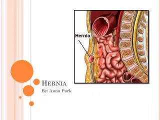
Hernia. Complications of herniorrhaphy are: 1 - wound sepsis 2 - Haematoma 3 - lymphocele (commoner after operations for femoral hernia); 4 - wound sinus (especially when foreign tissue is used for the repair); 5 - Division of spermatic cord (especially in infantile hernia operation)

E N D
Complications of herniorrhaphyare: 1 - wound sepsis 2 - Haematoma 3 - lymphocele (commoner after operations for femoral hernia); 4 - wound sinus (especially when foreign tissue is used for the repair); 5 - Division of spermatic cord (especially in infantile hernia operation) 6 - Testicular ischaemia (especially after large or recurrent hernia repairs) 7 - Testicular atrophy 8 – Hydrocele 9 - Nerve entrapment - Pain - Parasthesia or numbness;
10 - Recurrence (especially after operations for large . hernias in elderly males or sepsis) - 50% or recurrence happen within 2 years - False recurrrence ( new hernia ) 11 – general a - retention of urine b - respiratory complications c - thromboembolic complications Strangulated inguinal hernia : Strangulation of inguinal hernia can occurs at any time during life and in both sexes. Indirect inguinal hernias strangulate more common than the direct hernia due to wide neck in direct hernia. Sometimes a hernia strangulates on the first occasion that it descends; more often strangulation occurs in patients who have worn a truss for a long time, and in those with a partially reducible or irreducible hernia.
In order of frequency, the constricting agent is 1. the neck of the sac 2. the external abdominal ring (in children especially) 3. rarely adhesions within the sac. Contents: Usually small intestine is involved in the strangulation; the next most frequent content is omentum; sometimes both are implicated. For large intestine to become strangulated in an inguinal hernia is of the utmost rarity. Treatment : The treatment of strangulated hernia is by emergency operation. (‘The danger is in the delay, not in the operation) Vigorous resuscitation with intravenous fluids, NG tube aspiration and antibiotic is essential, also it is important to empty the bladder even by cath if necessary.
Inguinal herniotomyfor strangulation: An incision is made over the most prominent part of the swelling. The external oblique aponeurosis is exposed, and the sac, with its coverings, is seen issuing from the superficial inguinal ring. In all but very large hernias it is possible to deliver the body and fundus of the sac together with its coverings and (in the male) the testis onto the surface. Each layer covering the anterior surface of the body of the sac near the fundus is incised, and if possible it is stripped off the sac. The sac is then incised, the fluid there in is mopped up or aspirated very thoroughly, for it can be highly infected, The external oblique aponeurosis and the superficial inguinal ring are divided. Returning to the sac, a finger is passed into the opening, and employing the finger ass guide, the sac is slit along its length..
If the constriction lies at the superficial inguinal ring or in the inguinal canal, it is readily divided by this procedure. When the constricting agent is at the deep inguinal ring, by applying haemostats to the cut edge of the neck of the sac and drawing them downwards, and at the same time retracting the internal oblique upwards, it may be possible to continue slitting up the sac over the finger beyond the point of constriction. Then the neck ( deep ring ) of the sac is divided with a hernia knife in an upward and inward direction, i.e. parallel to the inferior epigastric artery. under vision. Once the constricting agent has been divided, the strangulated contents can be drawn down. Devitalisedomentum is excised after being securely ligated. Viable intestine is returned to the peritoneal cavity. Doubtfully viable and gangrenous intestine is dealt with. If the hernial sac is of moderate size and can be separated easily from its coverings, it is excised and dosed by a purse-string suture
Conservative measures : These are only indicated in infants. Children are given a sedative and then elevation of bed foot for no longer than 3 hours. In 75 per cent of cases reduction is effected and there appears to be no danger of gangrenous intestine being reduced. Manual reduction of irreducible and strangulated hernias under sedation is permissible in infants in whom the risk of intestinal gangrene appears to be almost nonexistent for many hours. For all other cases, vigorous manipulation (taxis) has no place in modern surgery, and is mentioned only to be condemned. Its dangers indude : 1 - Contusion or rupture of the intestinal wall.
2 - Reduction-en-masse. ‘The sac together with its contents, is pushed forcibly back into the abdomen; and as the bowel will still be strangulated by the neck of the sac, the symptoms are in no way relieved. 3 - Reduction into a loculus of the sac. 4 - The sac may rupture at its neck and its contents are reduced, not into the peritoneal cavity, but extraperitoneally. .
Sliding hernia : As a result of slipping of the posterior parietal peritoneum on the underlying retroperitoneal structures, the posterior wall of the sac is not formed of peritoneum alone, but by the sigmoid colon and its mesentery on the left, the caecum on the right and, sometimes, on either side by a portion of the bladder. It should be clearly understood that the caecum, appendix, or a portion of the colon wholly within a hernial sac does not constitute a sliding hernia. A small-bowel sliding hernia occurs once in 2000 cases; a sacless sliding hernia once in 8000 cases.
Clinical features : A sliding hernia occurs almost exclusively in males. Five out of six sliding hernias are situated on the left side; bilateral sliding hernias are exceedingly rare. The patient is nearly always over 40, the incidence rising with the weight of years. There are no clinical findings that are pathognomonic of a sliding hernia, but it should be suspected in every large globular inguinal hernia descending well into the scrotum. Large intestine is commonly present in a sliding hernia (or caecum and appendix in a right-sided case). Occasionally large intestine is strangulated in a sliding hernia; more often non strangulated large intestine is present behind the sac containing strangulated small intestine.
Treatment : Asliding hernia is impossible to control with a truss, and as a rule the hernia is a cause of considerable discomfort. Consequently operation is indicated, and the results generally are good. Operation. It is unnecessary to remove any of the sliding hernial sac provided it is freed completely from the cord and the abdominal wall, and that it is replaced deep to the repaired fascia transversalis, In many instances it is desirable to perform orchiectomy in order to effect a secure repair. No attempt should be made to dissect the caecum or colon free from the peritoneum under the impression that these are adhesions, in which case peritonitis or a faecalflstula resulting from necrosis of a devascularised portion of the bowel may occur. his is specially liable to occur on the left side, as vessels in the mesocolon may be injured.
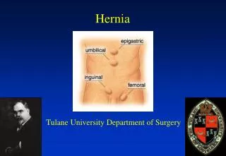
Femoral hernia Femoral hernia is the third most common type of hernia (incisional hernia comes second). It accounts for about 20 per cent of hernias in women, and 5 per cent in men. The overriding importance of femoral hernia lies in the facts that it cannot be controlled by a truss, and that of all hernias it is the most liable to become strangulated mainly because of the narrowness of the neck of the sac and the rigidity of thefemoral ring.
Femoral hernias ■ More common in women ■ Cannot be controlled with a truss ■ Have a high incidence of strangulation ■ Should be operated on as soon as possible
Surgical anatomy : The femoral canal occupies the most medial compartment of the femoral sheath, and it extends from the femoral ring above to the saphenous opening below. It is 1.25cm long, and 1.25 cm wide at its base, which is directed upwards. The femoral .canal contains fat, lymphatic vessels, and the lymph node of Cloquet. It is dosed above by the septum cru.rale, a condensation of extraperitoneal tissue pierced by lymphatic vessels, and below by the cribriform fascia.
The femoral ring is bounded: 1 - anteriorlyby the inguinal ligament 2 - posteriorlyby Astley Cooper’s (iliopectineal) ligament, the pubic bone, and the fascia over the pectineus muscle 3 - medially by the concave knife-like edge of lacunar ligament, which is also prolonged along the iliopectineal line as Astley Cooper’s ligament 4 - laterally by a thin septum separating it from the femoral vein.
Sex incidence: The female to male ratio is about 2:1, but it is interesting that whereas the female patients are frequently elderly, the male patients are usually between 30 and 45 years. The condition is more prevalent in women who have borne children than in nulliparas. The broader female pelvis also predisposes to the condition. Pathology: A hernia passing down the femoral canal descends vertically asfar as the saphenous opening. While it is confined to the inelastic walls of the femoral canal the hernia is necessarily narrow, but once it escapes through the saphenous opening into the loose areolar tissue of the groin, it expands. sometimes considerably. A fully distended femoral hernia and its bulbous extremity may be above the inguinal ligament. By the time the contents have pursued so tortuous a path they are usually irreducible and apt to strangulate, a circumstance which is also favoured by the rigidity of the surrounds of the femoral ring.
Clinical features: Femoral hernia is rare before puberty. Between 20 and 40 years of age the prevalence rises, and continues to old age. The right side is affected twice as often as the left, and in 20 per cent of cases the condition is bilateral. The symptoms to which a femoral hernia gives rise are less pronounced than those of an inguinal hernia; indeed, a small femoral hernia may be unnoticed by the patient or disregarded for years, until perhaps the day it strangulates. Adherence of greater omentum sometimes causes a dragging pain. Rarely, a large sac is present.
Differential diagnosis: A femoral hernia has to be distinguished from the following. 1 - An inguinal hernia. An inguinal hernia lies above and medial to the medial end of the inguinal ligament at its attachment to the pubic tubere. The femoral hernia lies below this. Occasionally the fundus of a femoral hernia sac overlies the inguinal ligament. 2 - A saphenavarix. A saphenavarix is a saccular enlargement of the termination of the long saphenous vein and it is usually accompanied by other signs of varicose veins. The swelling disappears completely when the patient lies down, while a femoral hernia sac usually is still palpable. In both there is an impulse on coughing. A saphenavarix will, however, impart a fluid thrill to the examining fingers when the patient coughs, or when the saphenous vein below the varix is tapped with the fingers of the other hand. Sometimes a venous hum can be heard when a stethoscope is applied over a saphenavarix.
3 -An enlarged femoral lymph node. If there are other enlarged lymph nodes in the region the diagnosis is tolerably simple, but when Cloquet’s lymph node alone is affected the diagnosis may be impossible unless there is a due, such as an infected wound or abrasion on the corresponding limb or on the perineum. Doubt should be removed by immediate surgery. 4 -Lipoma. 5 -A femoral aneurysm. . 6 -A psouasabsccss. There is often a suprainguinal fluctuating swelling — an iliac abscess — . Examination of the spine and a radiograph will settle the diagnosis. 7 - A distended psoas bursa. The swelling diminishes when the hip is flexed, and osteoarthrosis of the hip is present. 8 -Rupture of the adductor longsswith haematoma.
Strangulated femoral hernia : A femoral hernia strangulates frequently and gangrene develops rapidly. This is accounted for by the narrow, unyielding femoral ring. In 40 per cent of cases the obstructing agent is not lacunar ligament but the narrow neck of the femoral sac itself. The frequent occurrence of a Richter’s hernia also must be stressed. Treatment of femoral hernia: The constant risk of strangulation is sufficient reason for urging operation. A truss is contraindicated because of this risk.
:Umbilical hernia Exomphalos (syn. omphalocele) occurs once in every 6000 births; it is due to failure of all or part of the midgut to return to the coelom during early fetal life. Sometimes a large sac ruptures during birth. When the sac remains unruptured, it is semitranslucent and although very thin it consists of three layers — an outer layer of amniotic membrane, a middle layer of Wharton’s jelly, and an inner layer of peritoneum. There are two varieties of exomphalos: Exomphalos minor:The sac is relatively small and to its summit is attached the umbilical cord, Inadvertently sloop of small intestine or a Meckelsdiverticulum can be included in the ligature applied to the base of an umbilical cord containing this protrusion. Exomphalos major: The umbilical cord is attached to the inferior aspect of the swelling, which contains small and large intestine, and ,nearly always a portion of the liver, Half the cases belong to this group.
Treatment: Exomphalos minor. It is necessary only to twist the cord, so as to reduce the contents of the sac through the narrow umbilical opening into the peritoneal cavity, and to retain them by firm strapping. Despite a seropurulent discharge on no account must the strapping be removed for fourteen days. Exomphalos major. Operation within the first few hours of life is the only hope, otherwise the sac will burst. To prevent further distension of the contents of the sac, the infant should not be fed. A few newborn infants with a ruptured sac have survived following immediate operation and antibiotic therapy.
Umbilical hernia of infants and children. This is a hernia through a weak umbilical scar. The ratio of males to females is 2:1. Most do not become obvious until the infant is several weeks old. The hernia is often symptomless, but increase in the size of the hernia on crying causes pain, which makes the infant cry more. Small hernias are spherical; those that increase in size tend to assume a conical shape and are present apart from crying. Obstruction or strangulation below the age of three years is extremely uncommon Treatment: Conservative treatment is successful in about 95 per cent of cases. When the hernia is symptomless, reassurances of the parents is all that is necessary, for in a very high percentage of cases the hernia will be found to disappear spontaneously during the first few months of life. Cure may also be hastened by pulling the skin and abdominal musculature together by adhesive strapping placed across the abdomen. • .
Henuiorrhaphy. If the hernia persists at 2 years of age or older it is unlikely to resolve and herniorrhaphy is indicated.