
The Microscope
The Microscope . Unit 1: Biology I Fall. History of the Microscope. The Hand Lens has been used for over 600 years It was the first microscope used It allowed people to magnify text and objects. First Compound Microscope. Developed in 1595, by the Janssen brothers and Galileo

The Microscope
E N D
Presentation Transcript
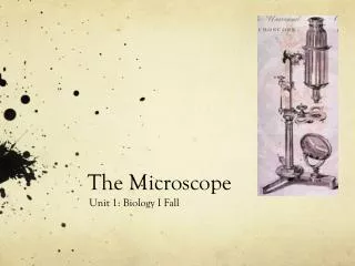
The Microscope Unit 1: Biology I Fall
History of the Microscope • The Hand Lens has been used for over 600 years • It was the first microscope used • It allowed people to magnify text and objects
First Compound Microscope • Developed in 1595, by the Janssen brothers and Galileo • Problem: images were blurred and had colored halos
The Father of Microscopy Anthony von Leeuwenhoek • Apprentice in a dry goods store • First to see and describe • Bacteria • Yeast plants • The teeming life in a drop of water • The circulation of blood corpuscles in capillaries.
Simple Glass Magnifiers • More than 500 years ago. • In 1600s, this “simple microscope” allowed scientists to see cells and bacteria • Problem: not enough magnification
Simple Compound Microscope • Invented in 1660s by Robert Hooke • Problem: all images had red or blue “halos” around them
Modern Compound Microscope • 1900s, started using iron instead of brass (cheaper) • Only one eyepiece (monocular) • Outside light source reflected onto mirror • Very functional • Still used today
Base • The bottom of the microscope, used for support • Hold this part with one hand when carrying a microscope ←
Mirror • Reflects the light so the specimen is easier to see ←
Stage • The flat platform where you place your slides. • It has a hole in it so light can shine through ←
Clip • Shiny clips on the top of the stage • Holds a slide in place ←
Arm • Supports the tube and connects it to the base • The part you hold when you carry the microscope ←
Coarse Adjustment • Large, round knob on the side of the microscope • Either moves the stage or the top part of the microscope up and down →
Fine Adjustment • Small, round knob on the side of the microscope • Used to fine tune the focus after using the coarse adjustment knob. →
Eyepiece → • The lens at the top that you look through. • 10X power
Body Tube • The long tube that holds the eyepiece and connects the objective →
Nosepiece • Rotating part of the microscope at the bottom of the body tube. • It holds the objective lenses ←
High Power Objective • The longest objective lens • The highest magnification • 40X lens (40X x10X = 400X magnification) →
Low Power Objective • The shortest objective lens • The lowest magnification • 4X lens (4X x10X = 40X magnification) ←
Mid Power Objective • The medium length objective lens • The medium magnification • 10X lens (10X x10X = 100X magnification) ←
Diaphragm • Controls the amount of light going through the hole in the stage →
Eyepiece BodyTube RevolvingNosepiece Arm ObjectiveLens Stage StageClips CoarseFocus Diaphragm FineFocus Light Base


