Protocol for managing para -pneumonic effusions
290 likes | 490 Views
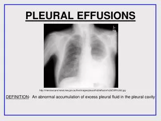
Antigona Trofor U.M.P.”Gr. T. Popa ” Iasi. Protocol for managing para -pneumonic effusions. When..plan. A. Investigate Pleural effusion. A. A. Work-up of pleuritis. Proteins > 35 G/l Light’s criteria PF/S protein >0.5 PF/S LDH >0.6 PF LDH > 2/3 of the upper limit of normal.

Protocol for managing para -pneumonic effusions
E N D
Presentation Transcript
AntigonaTrofor U.M.P.”Gr. T. Popa” Iasi Protocol for managing para-pneumonic effusions
When..plan A InvestigatePleural effusion
A A
Work-up of pleuritis • Proteins > 35 G/l • Light’s criteria • PF/S protein >0.5 • PF/S LDH >0.6 • PF LDH > 2/3 of the upper limit of normal
Work-up of pleuritis • Cytology is non-diagnostic in 40% • Pulmonary embolism ? • Malignancy • Tuberculosis • Idiopathic effusion
Exudative pleural effusions pH< 7.30 (or glucose < 60 mg/dL) Diagnosis is limited to 6 causes: Empyema Malignancy Esophageal rupture Tuberculous pleurisy Lupus pleurisy Rheumatic pleurisy Empyema
Eosinophilic pleuritis(pf eosin./total nucl. pf cells>10%) • Pneumothorax • Hematothorax • Drug reactions • Benign asbestos pleuritis • Lymphoma, carcinoma • Churg-Strauss syndrome • Infections (fungal, parasitic) • > 10% eosinophils rules out tuberculosis!
> 80% lymphocytes in pf • Tuberculosis • Chylothorax • Lymphoma • Trapped lung • Sarcoidosis • Chronic Rheumatic pleuritis • Yellow nail syndrome • Post- coronary artery by pass
Tuberculous pleuritis • PPD may be negative in 30% of cases • 12% in HIV negative patients • 47% in HIV positive patients • Eosinophils > 10% rule out tuberculosis • Mesothelial cells > 5% rule out TBC • Pleural fluid TB culture may be positive in only 20%
Why should you perform thoracoscopy ?(Pleural effusions) • Thoracocenthesis • Non-diagnostic in 20-60% • False-negative for malignancy • Specific diagnosis rare
Work-up of pleuritis • Blind pleural biopsy should only be performed in areas with high incidence of tuberculosis (in resource-poor countries) Diacon et al. Eur Resp J 2003;22: 589-91 Light RW. J Bronchology 1998;5:332-336
When ..plan B Investigatepneumonia
B Para-pneumonic effusion (PPE)? • Pleural effusion complicate 20-57% of hospitalized pneumonias • Depending on responsible organism for pneumonia • Any pneumonia should be assessed for para-pneumonic effusion • If more than minimal effusion, pleural fluid needs to be sampled.
Factors Associated with Poor Prognosis( require invasive procedures) • 1. Pleural fluid is pus • 2. Pleural fluid bacterial smears are positive • 3. Pleural fluid glucose is less than 60 mg/dl • 4. Pleural fluid bacterial cultures are positive • 5. Pleural fluid pH is less than 7.20 • 6. Pleural fluid LDH is more then three times the upper limit of normal • 7. Pleural fluid is loculated
Categorizing Risk for Poor Outcome in Patients With PPE Colice GLet al. Medical and surgical treatment ofparapneumonic effusions: An evidence-based guideline, Chest, 2000
Pleural fluid sampling • Diagnostic thoracentesis • Therapeutic thoracentesis • Insertion of small chest tube Wait and see
Treatment goals and options in pleural infection Loddenkemper R. et. al, Eur.Respir.Mon.,2004
Fibrinolytics? • “It is my recommendation that fibrinolytics should be reserved for patients in centers without access to video assisted thoracic surgery (VATS) and for patients who are not surgical candidates.” * * R.Light, 2008
Antibiotic therapy Loddenkemper R. et. al, Eur.Respir.Mon.,2004
Local pleural treatment options Loddenkemper R. et. al, Eur.Respir.Mon.,2004 Treatment Options
Procedural approach in the local treatment ofempyema (Lungenklinik Heckeshorn) Medical toracoscopy Drainage: Toracoscopic/image-guided double-lumen trocar catheter insertion, 20–28F, length 40 cm. Irrigation: 1,000 mL normal saline solution20 mL 2% povidone iodine,until clear irrigationfluid recovered. Instillation (fibrinolysis) 200,000 IU streptokinase50,000 IU streptodornase, tube clamped 4–8 h (tolerancedependent). Duration14 days Side effects Fever> 38 C (42%), pain (10%) Precautions Postural maneuvers (diseased side in dependent position), no bronchial- pleural fistula, no allergy.
Medical or surgical treatment in PPE and empyema? Loddenkemper R. et. al, Eur.Respir.Mon.,2004
Features suggesting additional local treatment in para-pneumonic effusions Loddenkemper R. et. al, Eur.Respir.Mon.,2004
Loculated pleural effusions Options? • Insertion of multiple chest tubes • Intrapleural administration of fibrinolitics • Thoracoscopy • Decortication • Open drainage procedure VATS is prefered
Evidence based guidelines of PPE treatment (ACCP) * Colice et. all, Chest, 2000 PPE should be considered in all pneumonia (C) Drainage of PPE should be based on estimated poor outcome risk (D) Risk 1 and 2 may not require drainage (D) Drainage recommended in risk 3 , 4 category (D) Therapeutic thoracentesis or tube thoracostomy alone appears insufficient in most of risk 3, 4 (C); reevaluation after several hours is useful (D) Fibrinolytics, VATS and surgery are acceptable approaches in risk 3, 4 categories (C)
TREATMENT OF PARAPNEUMONIC PLEURAL EFFUSION AND EMPYEMA DIAGNOSIS (Etiology, stage, complications) Exudative stage Fibrinopurulent stage Organisational stage complications Other causes Additional local treatment Antibiotics only Surgery (VATS or thoracotomy) Drainage +/-Pleuroscopy +fibrinolytics +irrigation Successful No success Continue No success