Download
1 / 42
420 likes | 442 Views
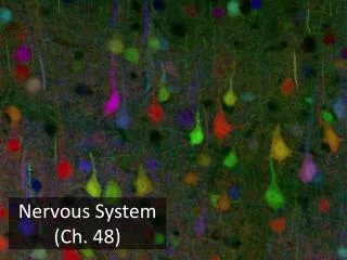
Learn about the key structures and functions of the nervous system, including neurons, neuroglial cells, neurotransmitters, and impulse conduction. This review covers various classifications and processes crucial to understanding neural activity.

E N D
AnswErs CH. 10 REVIEW INTRO TO NERVOUS SYSTEM
1. Name these “branches”. dendrites
2. Name this part (red bracket). Cell body
3. Name the “dots”. Nissl bodies
4. Name this region outlined by yellow. Axon hillock
5. Name the part indicated by arrows on is outlined in yellow. Myelin sheath or Schwann cells
8. Name the part shown in yellow. Synaptic knob
9. Name the white boxes shown by arrow. Protein channel for Na+
10. Name the part shown in yellow. Post synaptic neuron
11. The black triangles represent what? neurotransmitter
14. Give the structural classification of this neuron. bipolar
15. Give the structural classification of this neuron. unipolar
16. Give the structural classification of this neuron. multipolar
17. Name this CNS neuroglial cell. satellite
Schwann 18. Name this CNS neuroglial cell.
19. Name this PNS neuroglial cell. Oligodendrocyte
20. Name this PNS neuroglial cell. Astrocyte
21. Name this PNS neuroglial cell. ependymal
22. Name this PNS neuroglial cell. microglia
23. Name the purple ions. Na+ -70
A = fastest D = slowest A B C D E 24. Choose the diagram with the fastest action potential speed and the slowest action potential speed.
25. Name the orange ions. K+ -70
26. Name the blue ions. Protein organic anions -70
27. Which letter represents a sodium potassium pump? B -70 A B C
depolarization 28. What does this region (bracket) represent? (hint: ends in –ation)
repolarization 29. What does this region (bracket) represent? (hint: ends in –ation)
31. Name these “indentations”. Nodes of Ranvier
32. What type of nerve impulse conduction is shown here? Saltatory conduction
33. What ion diffuses in here and allows the vesicles to undergo exocytosis and dump the neurotransmitters into the synapse? Calcium = Ca+
34. Look at the part encircled in the yellow dotted line. What is occurring here? reuptake
List the effectors for each yellowbox. Skeletal muscle Smooth muscle Cardiac muscle Glands
Choose the step this picture illustrates: • Depolarization • Hyperpolarization • Resting hyperpolarization
38. What does # 5 represent? hyperpolarization
39. What is the name for this structure shown by yellow arrow? Ligand voltage gated channel
40. In this diagram what is the specific role of Ach? It is the ligand