Download
1 / 10
100 likes | 504 Views
Computational requirements for Electrical Impedance Tomography of brain function. David Holder Medical Physics, UCL Clinical Neurophysiology, UCH. Research Clinical. Clinical Neurophysiology, UCH. Medical Physics, UCL David Holder Kirill Aristovich James Avery Tugba Doru Hwan Koo

E N D
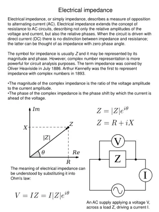
Computational requirements for Electrical Impedance Tomography of brain function David Holder Medical Physics, UCL Clinical Neurophysiology, UCH
Research Clinical Clinical Neurophysiology, UCH Medical Physics, UCL David Holder Kirill Aristovich James Avery Tugba Doru Hwan Koo Markus Jehl Mohamed Koronfel Elliott Magee Emma Malone Brett Packham Gustavo Santos Anna Vongerichten Collaborators Simon Arridge, CS, UCL Timo Betcke, Maths, UCL Ben Hanson, ME, UCL Paul Shearing, Chem Eng, UCL Eung-Je Woo, BME, S Korea GE Global Research, Schenectady, USA
Principal research :Electrical Impedance Tomography of brain function • A new medical imaging method • Ring(s) of electrodes are placed around the subject • A tiny current is passed at ~50 kHz between various combinations of electrodes • Images of impedance are produced ~10 times per second • Neuro applications : • Fast neural activity • Acute stroke • Epileptic seizures Plane 4 axilla Plane 3 Plane 2 Plane 1 xiphoid process electrodes 0 Frequency 1 MHz
Current development Images : actual EIT • EIT of brain function works in : • Tanks • Animal studies • Stroke • Epilepsy • Fast neural • Evoked responses • Does not yet work in patients Perspex rod
Long term goal – fast neural activity, 1 ms, 1 mm Every millisecond
Recording protocol (illustrated for square wave) EP 17 11 Voltage 25 Square wave Evoked potential Impedance change 12 3 23 5 + 27 16 1 29 21 7 + 26 13 2 I 28 20 8 24 15 4 19 9 = 22 14 6 Voltage 18 10 Record V 500 ms Oh, T., Gilad, O., Ghosh, A., Schuettler, M., Holder, D. S. (2011). A novel method for recording neuronal depolarization with recording at 125-825 Hz: implications for imaging fast neural activity in the brain with electrical impedance tomography. Med Biol Eng Comput 49(5), 593-604
Imaging 17 11 25 12 3 30x ~40 min 23 5 I 16 1 29 21 7 26 13 2 28 8 1min (120 av) 1min (120 av) 1min (120 av) 20 24 15 4 19 9 22 14 6 0.2mm, 662,788 elements Problem 18 Inverse 10 Adaptive, 359,797 elements Problem Forward 0.8mm, 49,973 elements Image ~300um, τ 8 ms reconstruction 1M element rat brain FEM mesh
Computational considerations Forward Modelling • Forward solution: • Computational complexity: • O(N); O(NlogN); • 2.106 - 100.106 FEM mesh • → ~ 10-100 Gb Memory • ~ 1-30 Gigaflops • ~ 10min - 4 hours
Image reconstruction • Total variation (TV) regularization: • Validation on saline tank phantom using 75k-element cylindrical mesh
Projection for future • Before optimization : • Practical • ? 10.106 FEM mesh → ~ 100 Gb Memory ~ 100 Gigaflops ~ 2 days per image • Ideal • ? 100.106 FEM mesh → ~ 2 Tb Memory ~ 1 Teraflop ~ 20 days per image • Optimized • ? 106 FEM mesh → ~ 10 Gb Memory ~ 1 Gigaflop ~ 10s per image