Download

1 / 18
180 likes | 477 Views
WANDERING OVARY. DR. J.CHITRA M.D , DGO, D.NB ASST PROFESSOR KANYAKUMARI GOVT MEDICAL COLLEGE. Clinical history. 24 years old young unmarried girl Acute abdominal pain for 4 days Attained menarche 14 th year Periods once in 2 months / 3 days Painless, scanty periods.

E N D
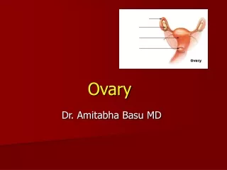
WANDERING OVARY DR. J.CHITRA M.D , DGO, D.NB ASST PROFESSOR KANYAKUMARI GOVT MEDICAL COLLEGE
Clinical history • 24 years old young unmarried girl • Acute abdominal pain for 4 days • Attained menarche 14th year • Periods once in 2 months / 3 days • Painless, scanty periods
Previous history • 1 year back, was treated for the same acute abdominal pain elsewhere. • Diagnosed as bilateral multiloculated ovarian cysts • Advised surgery- she refused • Treated conservatively • She was symptom free for one year
Scan report • Scan taken on 5.10.04 • Rt.Ovary 12x12cm Cystic, multilocular, thin walled • Lt Ovary 11x12 cm, cystic
CLINICAL EXAMINATION • O/E - P/A no mass palpable • P/R - Cystic, tender mass felt occupying the pouch of Douglas • Routine investigations normal • Ultrasonagram • Uterus normal • right ovarian cyst –multi loculated 12 x 12 cm • Left ovary –not visualized
LAPAROSCOPY FINDINGS • Right Ovary • Twisted • Cystic, Multilocular • Variable in size • Thin walled
Procedure on Right ovarian cyst • Untwisting of the ovarian cyst done • Cystectomy done • Remaining ovarian tissue left out LEFT OVARY NOT SEEN AT ITS SITE
Amputated site of left ovary OVARY ATTACHED TO OMENTUM
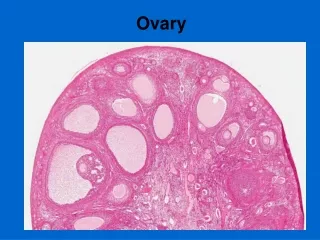
WANDERING OVARY • Normal looking ovary ,3.5 cm x 3 cm x 2 cm in size, seen attached to omentum • Feeding vessels are seen from omentum
Procedure on Right ovarian cyst • Untwisting of the ovarian cyst done • Cystectomy done • Remaining ovarian tissue left out LEFT OVARY NOT SEEN AT ITS SITE
HPE SIMPLE SEROUS CYST RIGHT OVARIAN CYST • AMPUTED OVARIAN BIOPSY CONFIRMED OVARIAN TISSUE – HPE
FOLLOW UP • REGULAR MENSTRUATION STARTED AFTER THREE MONTHS • AFTER ONE YEAR ULTRA SOUND REVEALED NORMAL UTERUS. RIGHT OVARY 4 x 3cms LEFT OVARY NOT SEEN • NO OTHER CYSTIC MASS SEEN • PLAIN X-RAY – NO CALCIFICATION NOTED
THANK YOU THANK YOU