Normal Hemostasis
440 likes | 1.32k Views
Normal Hemostasis. Galila Zaher Consultant Hematologist KAUH. Function of Haemostasis. Prevention of blood loss from intact vessels Arrest of bleeding from damaged vessels Blood vessel reaction to injury Platelet plug formation at site of damage. BLOOD CLOTTING.

Normal Hemostasis
E N D
Presentation Transcript
Normal Hemostasis Galila Zaher Consultant Hematologist KAUH
Function of Haemostasis • Prevention of blood loss from intact vessels • Arrest of bleeding from damaged vessels • Blood vessel reaction to injury • Platelet plug formation at site of damage
BLOOD CLOTTING Plasma protein clotting factors Vascular endothelium Platelets
COAGULOPATHIES Bleeding Thrombosis Clotting factors Natural anticoagulant platelets
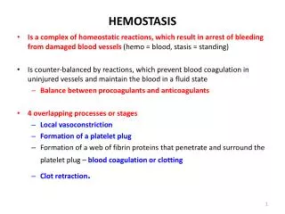
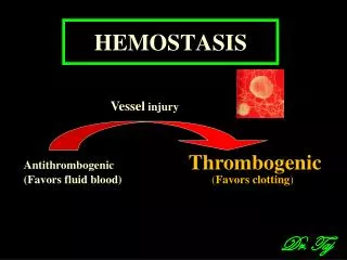
Vessel wall Platelet Blood coagulation Fibrinolytic system Inhibitors Local vasoconstriction (noradrenaline & serotonin) Platelet release thromboxane A2 (a vasoconstrictor) Prostacyclin is released counters effects of thromboxane A2 Normal Hemostasis
Vessel wall Platelet Blood coagulation Fibrinolytic system Inhibitors Adhesion Shape change Aggregation Release Reaction Normal Hemostasis
ADP Aggregation Aggregation Aggregation GpIIb/IIIa GpIIb/IIIa GpIIb/IIIa GpIIb/IIIa GpIIb/IIIa GpIIb/IIIa Adrenaline Adhesion Adhesion vWF Endothelium Exposed Collagen Platelet Activation COLLAGEN THROMBIN ADP GpIIb/IIIa Platelet GpIb Adrenaline Adhesion
Platelet activation Primary hemostasis Count &function Immediate Fibrin generation Secondary hemostasis Plasma clotting factors Delayed Clot formation
VWF • The largest multimers of vWF greater adhesive & prothrombotic potential more sites to interact with: • Substrates in extra-cellular matrices and • Platelet receptors.
F.VIII:C F. VIIIC:Ag vWF:Ag vWF B:Co vWF R:Co
Functions of vWF • Platelet Adhesion (to extracellular matrices):GP Ib-IX-V complex • Platelet Adhesion to each other (Aggregation):Mediated by vWF & fibrinogen binding to:GPIb-IX-V & GP IIb-IIIa
Clotting factor production Liver: source of plasma clotting factors except VWF Factor VIII: produced by liver & endothelium VWF: endothelial cells & megakaryocytes Vitamin K dependent clotting factors are: II, VII, IX, X
COAGULATION PATHWAYS Intrinsic & extrinsic pathways “conclude” in the common pathway Intrinsic pathway clotting factors Extrinsic pathway clotting factors Common pathway clotting factors
Intrinsic Pathway All clotting factors are within the blood vessels Clotting slower Activated partial thromboplastin test (aPTT) Extrinsic Pathway Initiating factor is outside blood vessels - tissue factor Clotting faster Prothrombin test (PT)
The “Cascade”, “Waterfall” model: • Macfarlane (1964), Davie & Ratnoff (1964)} • Drawbacks of the ‘cascade model’: • Model inadequate to explain pathways leading to hemostasis in vivo • Deficiency of FXII, HMK,PK does not cause bleeding • Activation of FX by the extrinsic pathway does not compensate for the lack of FVIII or FIX in hemophiliacs
Initiation TF VIIa IXa Fibroblast Propagation Xa Prothrombin Thrombin Thrombin Xa IXa XIa Prothrombin XIa VIIIa Platelet Activated platelets Amplification
Alternative (Cell-based) Model • The initiation phase: Commences on TF-bearing cells (fibroblasts) FXa, IXa & thrombin Initiation of the coagulation process • The amplification phase: Coagulation moves from TF-bearing cells to activated platelets, • The propagation phase: The active proteases combine with cofactors on platelet surface to generate thrombin; End-result is fibrin polymerization
Vessel wall Platelet Blood coagulation Fibrinolytic system Inhibitors Fibrinolysis Fibrin is digested by enzymes from plasma and from cells. Endothelium replaces the fibrin
Plasminogen TPA Thrombin XIIa • Plasmin
Activation of fibrinolysis damaged cells thrombin inflammation mental/physical stress trauma PAI t-PA extrinsic pathway plasminogen plasmin antiplasmin cross-linked fibrin fibrinogen X-FDP (D-Dimer, cross-linked oligomers, DD/E ...) FDP (X,Y,D,E)
Generation Of Fibrin and D-Dimer D D E D D E E E E E D D D D D D D D D D D D E E E E E E E E E D D D D D D D D E E E E E E E E fibrinogen E thrombin fibrin FpA, FpB fibrin polymer F XIIIa cross-linked fibrin (clot) D-dimer cross-linkage
COAGLATION INHIBITORS • Antithrombin (FXIa, IXa, Xa & IIa) • Protein C/S complex (F Va & F VIIIa) • Tissue factor pathway inhibitor -(TFPI)
XIIa XII XI XIa VIIa VII IX IXa X X II IIa Va Fibrinogen Fibrin ATIII Ps Xa VIIIa Pc PAI-1 Lysis of fibrin
Tissue factor +Factor VIIa TFPI Factor IXa -FVIIIa Factor Xa +FVa Anti Thrombin III Protein C Factor IIa (thrombin) Factor XIa Fibrinogen Fibrin
Extrinsic pathway VII + TF ----->VIIa/TF Intrinsic pathway XII ---> XIIa XI---------XIa • IX --------> IXa • + VIII APC PC +PS • Ca +PL • X----------------------> Xa [Common pathway] • V+Ca+PL • Prothrombin -------------> thrombin AT • v • fibrinogen--------------> fibrin