Hemostasis
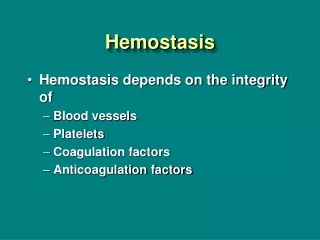
Hemostasis. Mechanisms of Hemostasis Hypercoagulability States Bleeding Disorders. hemostasis refers to the stoppage of blood flow

Hemostasis
E N D
Presentation Transcript
Mechanisms of Hemostasis Hypercoagulability States Bleeding Disorders
hemostasis refers to the stoppage of blood flow The normal process of hemostasis is regulated by a complex array of activators and inhibitors that maintain blood fluidity and prevent blood from leaving the vascular compartment.
Hemostasis is normal when it seals a blood vessel to prevent blood loss and hemorrhage. It is abnormal when it causes inappropriate blood clotting or when clotting is insufficient to stop the flow of blood from the vascular compartment.
Disorders of hemostasis fall into two main categories: the inappropriate formation of clots within the vascular system (i.e., thrombosis) and the failure of blood to clot in response to an appropriate stimulus (i.e., bleeding)
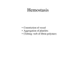
MECHANISMS OF HEMOSTASIS Hemostasis is divided into five stages: vessel spasm, formation of the platelet plug, blood coagulation or development of an insoluble fibrin clot, clot retraction, and clot dissolution
Vessel spasm Vessel spasm is initiated by endothelial injury and caused by local and humoral mechanisms. A spasm constricts the vessel and reduces blood flow. It is a transient event that usually lasts less than 1 minute. Thromboxane A2 (TXA2), a prostaglandin released from the platelets, contributes to the vasoconstriction. A second prostaglandin, prostacyclin, released from the vessel endothelium, produces vasodilation and inhibits platelet aggregation.
Formation of the Platelet Plug The platelet plug, the second line of defense, is initiated as platelets come in contact with the vessel wall. The outside of the platelet membrane is coated with glycoproteins that repulse adherence to the normal vessel endothelium, while causing adherence to injured areas of the vessel wall, particularly the subendothelial layer.
The platelet membrane also has glycoprotein receptors that bind fibrinogen and link platelets together. Glycoprotein receptor antagonists have been developed and are selectively used in the treatment of acute coronary myocardial infarction
Platelet plug formation involves adhesion and aggregation of platelets. Platelet adhesion also requires a protein molecule called von Willebrand factor (vWF). vWF, which is produced by the endothelial cells of blood vessels, performs two important functions: it aids in platelet adhesion, and it circulates in the blood as a carrier protein for coagulation factor VIII.
Platelets are attracted to a damaged vessel wall, become activated, and change from smooth disks to spiny spheres, exposing receptors on their surfaces.
Adhesion to the vesselsubendothelial layer occurs when the platelet receptor bindsto vWF at the injury site, linking the platelet to exposed collagenfibers
Blood Coagulation • Blood coagulation is controlled by many substances that • promoteclotting (i.e., procoagulation factors) or • inhibit it (i.e., anticoagulationfactors). • The action of one coagulation factor orproenzyme is designed to activate the next factor in the sequence(i.e., cascade effect). • Abnormalitiesof the clotting process occur when one or more ofthe factors are deficient or when conditions lead to inappropriateactivation of any of the steps.
The chemical events in the blood coagulation processinvolve a number of essential steps that result in the conversionof fibrinogen, a circulating plasma protein, to the fibrinstrands that enmesh platelets, blood cells, and plasma to formthe clot
The initiation of the clotting process occursby way of the intrinsic or the extrinsic coagulation pathways
The intrinsic pathway, which is a relativelyslow process, begins in the blood itself. • The extrinsic pathway,which is a much faster process, begins with tissue or vesseltrauma and the subsequent release of a complex of several factors,called tissue factor, or tissue thromboplastin. • The terminalsteps in both pathways are the same: • the activation of factor X, • the conversion of prothrombin to thrombin, and • the conversionof fibrinogen to fibrin.
The intrinsic system is activated as blood comes in contactwith collagen in the injured vessel wall and the • extrinsicsystem when blood is exposed to tissue extracts.
With few exceptions, almost all theblood-clotting factorsare synthesized in the liver. • Vitamin K is required for the synthesisof prothrombin, factors VII, IX, X, and protein C. • Calcium(factor IV) is required in all but the first two steps of theclotting process.
Coagulation is regulated by several natural anticoagulants.Antithrombin III inactivates coagulation factors and neutralizesthrombin
Clot Retraction • After the clot has formed, clot retraction, which requires largenumbers of platelets, contributes to hemostasis by squeezingserum from the clot and joining the edges of the broken vessel.
Clot Dissolution • The dissolution of a blood clot begins shortly after its formation;this allows blood flow to be re-established and permanenttissue repair to take place • The process bywhich a blood clot dissolves is called fibrinolysis.
Plasminogen,the proenzyme for the fibrinolytic process, normally is presentin the blood in its inactive form. • It is converted to its activeform, plasmin, by plasminogen activators formed in the vascularendothelium, liver, and kidneys.
The plasmin formedfrom plasminogen digests the fibrin strands of the clotand certainclotting factors • Circulating plasmin is rapidlyinactivated by α2-plasmin inhibitor
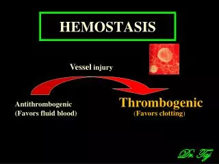
Hints on normal vascular hemostasis Normal hemostasis: results from well-regulated processes that maintain blood in a fluid, clot-free state in normal vessels while inducing the rapid formation of a localized hemostatic plug at the site of vascular injury. Thrombosis:is the pathologic converse to hemostasis it can be thought of as the formation of a blood clot (thrombus) in uninjured vessels, or thrombotic occlusion of a vessel after relatively minor injury. Both hemostasis and thrombosis are dependent on three general components: the vascular wall, platelets, and the coagulation cascade
Hints on normal vascular hemostasis • Normal hemostasis: results from well-regulated processes that maintain blood in a fluid, clot-free state in normal vessels while inducing the rapid formation of a localized hemostatic plug at the site of vascular injury. • Thrombosis:is the pathologic converse to hemostasis it can be thought of as the formation of a blood clot (thrombus) in uninjured vessels, or thrombotic occlusion of a vessel after relatively minor injury. • Both hemostasis and thrombosis are dependent on three general components: the vascular wall, platelets, and the coagulation cascade
Endothelial cells modulate several aspects of normal hemostasis: - On the one hand, at baseline they exhibit antiplatelet, anticoagulant, and fibrinolytic properties. - On the other hand, they are capable (after injury or activation) of exerting procoagulant functions
THROMBOSIS • Three primary influences predispose to thrombus formation, the so-called Virchow triad:
Endothelial injury • Physical loss of endothelium leads to: - exposure of subendothelial collagen (and other platelet activators) - adherence of platelets - release of tissue factor
Dysfunctional endothelium may elaborate greater amounts of procoagulant factors(e.g., adhesion molecules to bind platelets, tissue factor, PAI, etc.) and smaller amounts of anticoagulant effectors(e.g., thrombomodulin, PGI2, t-PA).
Abnormal blood flow • Turbulence contributes to arterial and cardiac thrombosis by causing endothelial injury or dysfunction • Stasis is a major factor in the development of venous thrombi
Normal blood flow is laminar such that the platelet elements flow centrally in the vessel lumen, separated from the endothelium by a slower-moving clear zone of plasma. Stasis and turbulence therefore: • a) Disrupt laminar flow & bring platelets into contact with the endothelium • b) Prevent dilution of activated clotting factors by fresh-flowing blood
c) Retard the inflow of clotting factor inhibitors and permit the build-up of thrombi • d) Promote endothelial cell activation, predisposing to local thrombosis, leukocyte adhesion, and a variety of other endothelial cell effects • Examples: Myocardial infarction
HYPERCOAGULABILITY STATES • There are two general forms of hypercoagulabilitystates: • conditions that create increased platelet function and • conditions that cause accelerated activity of the coagulationsystem.
Increased Platelet Function • The causes of increased platelet function are • disturbancesin flow, • endothelial damage, and • increased sensitivity of plateletsto factors that cause adhesiveness and aggregation
Increased Clotting Activity • results from factors that increase theactivation of the coagulation system, including stasis of bloodflow and alterations in the coagulation components of theblood (i.e., an increase in procoagulation factors or a decreasein anticoagulation factors)
Tips on thrombi • Thrombi may develop anywhere in the cardiovascular system: within the cardiac chambers, on valve cusps, or in arteries, veins, or capillaries. • They are of variable size and shape, depending on the site of origin and the circumstances leading to their development. • Arterial or cardiac thrombi usually begin at a site of endothelial injury or turbulence.
Venous thrombi characteristically occur in sites of stasis • The propagating tail may not be well attached and, particularly in veins, is prone to fragment, creating an embolus
Development of Coronary Atherosclerosis • Coronary atherosclerosis is an inflammatory disease characterized by the accumulation of white blood cells, cell debris, fatty substances (cholesterol and fatty acids), calcium, and fibrous tissue on the walls of the coronary arteries that supply the heart muscle. • As plaque slowly increase in size over many years, the artery narrows in places (stenosis), and blood flow to the heart is reduced.
BLEEDING DISORDERS • Bleeding disorders or impairment of blood coagulation can resultfrom defects in any of the factors that contribute to hemostasis. • Defects are associated with • platelets, • coagulation factors,and • vascular integrity.
Platelet Defects • Disorders of platelet plug formation include • a decreasein platelet numbers due to inadequateplatelet production (bone marrow dysfunction), • excess platelet destruction (thrombocytopenia), • abnormal platelet function (thrombocytopathia),or • defects in von Willebrand factor.
Coagulation Defects • Impairment of blood coagulation can result from deficienciesof one or more of the known clotting factors. • Deficiencies canarise because of • defective synthesis, • inherited defects, or • increasedconsumption of the clotting factors.
Bleeding thatresults from clotting factor deficiency typically occurs afterinjury or trauma. • Large bruises, hematomas, or prolongedbleeding into the gastrointestinal or urinary tracts or joints arecommon.
Coagulation Defects • Impaired Synthesis of Coagulation Factors • factors V, VII, IX, X, XI, and XII; prothrombin; andfibrinogen are synthesized in the liverIn liver disease, synthesisof these clotting factors is reduced • Hemophilia A • Deficiencyor defect in factor VIII • Von Willebrand Disease • Deficiencyof or defect in vWF • results in reduced plateletadhesion