Hemostasis
Hemostasis. ELV. Important points to be illustrated.

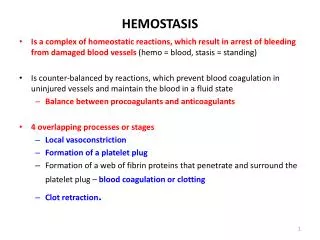
Hemostasis
E N D
Presentation Transcript
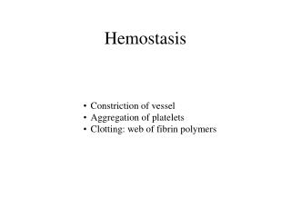
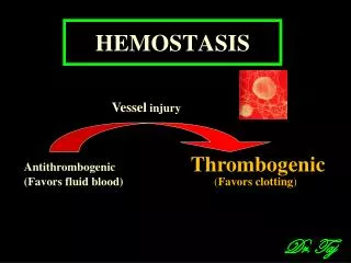
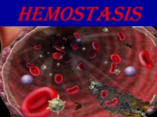
Important points to be illustrated • Endothelial cells in intact blood vessels inhibit coagulation by secreting tissue plasminogen activator (t-PA), thrombomodulin (inhibits thrombin) and by enzymatically inactivating adenosine diphosphate(ADP). Undamaged endothelium also inhibits platelet aggregation, by secreting nitric oxide and prostacyclin. If the endothelium is damaged, it stops secreting these inhibitors and instead secretes von Willebrandfactor (vWf) and tissue thromboplastin (TPL) which contribute to a repair process that involves platelet recruitment, activation, aggregation and thrombus formation (clotting). • Three mechanisms contribute to this repair process: • 1) Vascular spasm - damaged blood vessels constrict, slowing down the loss of blood and bringing circulating platelets and other factors into proximity with the damaged area. Shear stress is a major factor in clot formation. • 2) Platelet plug formation – circulating vWf binds to the vascular sub-epithelium. The platelet GP Ib-IX receptor adheres to vWf which recruits platelets. This initiation allows the platelet GP Ia-IIa receptor to bind sub-epithelial collagen, a stronger interaction. The platelet GPVI receptor also binds collagen and generates intracellular signals that ultimately bring about platelet activation. Part of this is GP IIb-IIIa activation, which allows it to bind fibrinogen. Bound fibrinogen molecules cross-link platelets into a plug (aggregation). Another consequence of platelet activation is a change of platelet shape from discoid to ‘spiky’ as they form pseudopdia which both cover the exposed subepithelial surface and aid the entrapment of circulating platelets. Activated platelets degranulate, releasing signals such as ADP which stimulate platelet activation, and thromboxane, serotonin and prostaglandin which maintain vasoconstriction. This lead to the recruitment and activation of further platelets. This primary hemostasis process quickly generates (20 s) a soft platelet plug consisting largely of platelets and fibrinogen. • 3) Blood coagulation - Clots form upon the conversion of fibrinogen to fibrin(secondary hemostasis). • I’ve not described the process of clot dissolution as you have already included that in the diagram. • This may be more details than necessary – please feel free to ask for clarifications