Download
1 / 63
630 likes | 1.08k Views
C H A P T E R T H I R T E E N. THE NERVOUS SYSTEM: NEURAL TISSUE. Two organ systems coordinate and direct activities of body. Nervous system Swift, brief responses to stimuli Endocrine system Adjusts metabolic operations Directs long-term changes.

E N D
C H A P T E R T H I R T E E N THE NERVOUS SYSTEM: NEURAL TISSUE
Two organ systems coordinate and direct activities of body • Nervous system • Swift, brief responses to stimuli • Endocrine system • Adjusts metabolic operations • Directs long-term changes
Nervous system includes all neural tissue in body • Central Nervous System • Brain and spinal cord • Peripheral Nervous System • All neural tissue outside CNS
Functional divisions of nervous system • Afferent • Sensory information from receptors to CNS • Efferent • Motor commands to muscles and glands • Somatic division • Voluntary control over skeletal muscle • Autonomic division • Involuntary regulation of smooth and cardiac muscle, glands
Cells in Nervous Tissue • Neurons • Neuroglia
Neuroglia (Glia) • about half the volume of cells in the CNS • smaller than neurons • 5 to 50 times more numerous • do NOT generate electrical impulses • divide by mitosis • Four types in the CNS • Astrocytes • Oligodendrocytes • Microglia • Ependymal cells
Astrocytes • Largest of glial cells • Most numerous • Star shaped with many processes projecting from the cell body • Help form and maintain blood-brain barrier • Provide structural support for neurons • Maintain the appropriate chemical environment for generation of nerve impulses/action potentials • Regulate nutrient concentrations for neuron survival • Regulate ion concentrations - generation of action potentials by neurons • Take up excess neurotransmitters • Assist in neuronal migration during brain development • Perform repairs to stabilize tissue
Oligodendrocytes • Most common glial cell type • Each forms myelin sheath around the axons of neurons in CNS • Analogous to Schwann cells of PNS • Form a supportive network around CNS neurons • fewer processes than astrocytes • round or oval cell body
Microglia • few processes • derived from mesodermal cells • that also give rise to monocytes • and macrophages • Small cells found near blood vessels • Phagocytic role - clear away dead cells • protect CNS from disease through phagocytosis of microbes • migrate to areas of injury where they clear away debris of injured cells - may also kill healthy cells
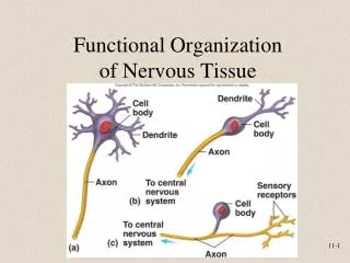
Representative Neuron http://www.horton.ednet.ns.ca/staff/selig/Activities/nervous/na1.htm • -neurofilaments or neurofibrils give cell shape and support - bundles of • intermediate filaments • -microtubules move material inside cell • -lipofuscin pigment clumps (harmless aging) - yellowish brown • 1. cell body or soma • -single nucleus with prominent nucleolus • -Nissl bodies • -rough ER & free ribosomes for protein synthesis • -proteins then replace neuronal cellular components for growth • and repair of damaged axons in the PNS
Neurons 2. Cell processes = dendrites (little trees) - the receiving or input portion of the neuron -short, tapering and highly branched -surfaces specialized for contact with other neurons -cytoplasm contains Nissl bodies & mitochondria
3. Cell processes = axons • Conduct impulses away from cell body-propagates nerve impulses to another neuron • Long, thin cylindrical process of cell • contains mitochondria, microtubules & neurofibrils - NO ER/NO protein synth. • joins the soma at a cone-shaped elevation = axon hillock • first part of the axon = initial segment • most impulses arise at the junction of the axon hillock and initial segment = trigger zone • cytoplasm = axoplasm • plasma membrane = axolemma • Side branches = collaterals arise from the axon • axon and collaterals end in fine processes called axon terminals • Swollen tips called synaptic end bulbs contain vesicles filled with neurotransmitters
http://bcs.whfreeman.com/thelifewire/content/chp44/4401s.swf Excitability • Ability of cell membrane to conduct electricity • Skeletal muscle fibers • Most neurons • Changes in membrane potential induces an action potential (AP) • the PM of neurons exhibits a membrane potential = electrical voltage difference across the membrane • in excitable cells like neurons this potential = resting potential • due to differences in sodium and potassium ion concentration in and out of the cell • potassium is higher inside cell, sodium is higher outside • inside of the cell has a higher concentration of negative phosphate ions and proteins = potential of -40 to -90 mV • the cell is said to be polarized • membrane has sodium/potassium pumps • to maintain specific concentrations of these ions in • & out of the neurons and therefore maintain the • resting membrane potential • -move three Na+ out and 2 K+ in • -inside of the neuron is slightly • negative
Action Potential • Resting membrane potential is -70mV • threshold usually -55 MV • Depolarization is the change from -70mV to +30 mV • Repolarization is the reversal from +30 mV back to -70 mV) http://www.blackwellpublishing.com/matthews/channel.html • action potential = nerve impulse • takes place in two stages: depolarizing phase (more positive) and repolarizing phase (more negative - back toward resting potential) • followed by a hyperpolarizing phase or refractory period in which no new AP can be generated
depolarization (increase in MP) results from opening of Na+ channels. Once threshold is reached, more Na+ channels open and a rapid increase in MP results outflow of K+ restores the MP. Na+ channels begin to open and K+ channels close. K+ outflow results in hyperpolarization (below resting) results in a refractory period. more slowly, depolarization also opens K+ channels which permit the outflow of K+ . The Na+ close the MP becomes more negative returning to resting
Local Anesthetics • Prevent opening of voltage-gated Na+ channels • Nerve impulses cannot pass the anesthetized region • Novocaine and lidocaine – blocks nerve impulses along nerves that detect pain
Synapse • Synapse • Site of intercellular communication between 2 neurons or between a neuron and an effector (e.g. muscle) • Originates in the soma • Travels along axons • Permit communication between neurons and other cells • Initiating neuron = presynaptic neuron • Receiving neuron = postsynaptic neuron • two types: chemical & electrical • NT will cause either and excitatory or inhibitory response • If the NT depolarizes the postsynaptic neuron= excitatory
Neurotransmitters • More than 100 identified • Some bind receptors and cause channels to open • Others bind receptors and result in a second messenger system • Results in either excitation or inhibition of the target • botulism causes paralysis through blockage of AcH release from motor • neurons • 1. small molecules: e.g. Acetylcholine (ACh) • -All neuromuscular junctions use ACh • -ACh also released at chemical synapses in the PNS and by some CNS neurons • -Can be excitatory at some synapses and inhibitory at others 2. Amino acids: glutamate & aspartate & GABA • Stimulate most excitatory neurons in the CNS (about ½ the neurons in the brain) • Binding of glutamate to receptors opens calcium channels = excitatory response • GABA (gamma amino-butyric acid) is an inhibitory neurotransmitter for 1/3 of all brain synapses Valium is a GABA agonist - enhancing its inhibitory effect
Neurotransmitters 3. Biogenic amines: modified amino acids • catecholamines:norepinephrine (NE), epinephrine, dopamine (tyrosine) • serotonin - concentrated in neurons found in the brain region = raphe nucleus • derived from trytophan • sensory perception, temperature regulation, mood control, appetite, sleep induction • feeling of well being • NE - role in arousal, awakening, deep sleep, regulating mood • epinephrine (adrenaline) - flight or fight response • dopamine - emotional responses and pleasure, decreases skeletal muscle tone • Parkinsons - muscle stiffness due to degeneration of dopanergic nerves • patients given L-Dopa (dopamine precursor) • amphetamines promote dopamine and NE release • isoproterenol binds to epinephrine receptors • - used in asthma to mimic the effects of epinephrine • schizophrenia - excess of dopamine • Zyprexa blocks dopamine and serotonin receptors • -antagonizes the effects of serotonin and dopamine • cocaine: blocks transporters for dopamine reuptake • Prozac, Paxil: blocks transporters for serotonin reuptake Other types: Nitric oxide - formed on demand in the neuron then release (brief lifespan) -role in memory and learning -produces vasodilation - Viagara enhances the effect of NO
Neuropeptides • widespread in both CNS and PNS • excitatory and inhibitory • act as hormones elsewhere in the body • -Substance P -- enhances our perception of pain • -opoid peptides: endorphins - release during stress, exercise • enkephalins - analgesics • (200x stronger than morphine) • -pain-relieving effect by blocking the release of • substance P • dynorphins - regulates pain and emotions • **acupuncture may produce loss of pain sensation because of release of opioid-like substances such as endorphins or dynorphins
Divisions of the nervous system • Sensory pathway • Ascending • Information from sensory receptors to CNS • Motor pathway • Descending • Information from CNS to skeletal muscle or glands • Direct pathways – cause precise, voluntary movements • Indirect pathways – result in involuntary movement (from brain stem)
Functional Classification of Neurons • Sensory (afferent) neurons • transport sensory information from skin, muscles, joints, sense organs & viscera to CNS • Motor (efferent) neurons • send motor nerve impulses to muscles & glands • Interneurons (association) neurons • connect sensory to motor neurons • 90% of neurons in the body
Sensory Neurons • Afferent division of PNS • Deliver sensory information from sensory receptors to CNS • free nerve endings: bare dendrites associated with pain, itching, tickling, heat and some touch sensations • Exteroceptors: located near or at body surface, provide information about external environment • Proprioceptors: located in inner ear, joints, tendons and muscles, provide information about body position, muscle length and tension, position of joints • Interoceptors: located in blood vessels, visceral organs and NS -provide information about internal environment -most impulses are not perceived – those that are, are interpreted as pain or pressure
Sensory Neurons • Sensory receptors cont… • mechanoreceptors: detect pressure, provide sensations of touch, pressure, vibration, proprioception, blood vessel stretch, hearing and equilibrium • thermoreceptors: detect changes in temperature • nociceptors: respond to stimuli resulting from damage (pain) • photoreceptors: light • osmoreceptors: detect changes in OP in body fluids • chemoreceptors: detect chemicals in mouth (taste), nose (smell) and body fluids -analgesia: relief from pain -drugs: aspirin, ibuprofen – block formation of prostaglandins that stimulate the nociceptors -novocaine – block nerve impulses along pain nerves -morphine, opium & derivatives (codeine) – pain is felt but not perceived in brain (blocks morphine and opiate receptors in pain centers)
Motor Neurons • Efferent pathways • Stimulate peripheral structures • Somatic motor neurons • Innervate skeletal muscle • Visceral motor neurons • Innervate all other peripheral effectors • Preganglionic and postganglionic neurons
Somatic nervous system (SNS): 1. sensory - neurons that convey sensory information from somatic receptors in the head, body wall, senses - to the CNS 2. control of motor output - neurons that conduct voluntary impulses to skeletal muscles -contributions from the basal ganglia, cerebellum, brain stem and SC 3. one neuron pathway – somatic motor neurons synapse directly with the effector 4. neurotransmitter – usually acetylcholine 5. effectors – skeletal muscles 6. responses - contraction
Autonomic nervous system (ANS): • sensory - neurons that convey info from autonomic sensory receptors in the visceral organs - to the CNS • 2. control of motor output - neurons that conduct impulses from the CNS to • smooth and cardiac muscle & glands • 3. two neuron pathway – preganglionic neurons extend from CNS and synapse with postganglionic neurons in an autonomic ganglion, postganglionic neurons then synapse with the effector • 4. neurotransmitter – preganglionic – ACh • -postganglionic – ACh or norepinephrine • 5. effectors – smooth & cardiac muscle, glands, • 6. responses – contraction or relaxation of SM • -increased or decrease heart contraction • -increased or decreased gland secretions
- motor output branch has two divisions: 1. sympathetic 2. parasympathetic -most organs are innervated by both divisions which have opposing functions e.g. sympathetic – increases heart rate parasympathetic – decreases rate -
Major Regions of the Brain Figure 15.1 Major Divisions of the Brain http://www.wisc-online.com/objects/framz.asp?objID=OTA502
I - Olfactory II - Optic III - Oculomotor IV-Trochlear V - Trigeminal VI - Abducens VII - Facial VIII - Acoustic IX - Glossopharyngeal X - Vagus XI - Accessory XII - Hypoglossal • -cranial nerves – 12 pairs • -considered part of the peripheral nervous system (PNS) • -olfactory & optic contain only sensory axons = sensory nerves • -some are motor nerves – e.g. oculomotor, trochlear etc…. • remaining are mixed nerves – both motor and sensory axons • “some say my mother bought my brother some bitter beer, my, my” http://www.wisc-online.com/objects/index.asp?objID=AP11504
The Olfactory Nerve (I) • Carries sensory information • Sense of smell from nasal mucosa to brain • Branches enter skull through cribiform plate • Synapses within olfactory bulbs
The optic nerve (II) (sensory) • Carries visual information • enters skull through optic canal of the sphenoid -right and left join at the optic chiasma (site of cross-over) -continue to brain as optic tracts
The oculomotor nerve (III) [Motor] • Primary source of innervation for extra-ocular muscles • also carries postganglionic fibers that innervate the ciliary muscles (lens shape) • exits through superior orbital fissure • The trochlear nerve (IV) [Motor] • Smallest cranial nerve • Innervates superior oblique eye muscle • also provides proprioception info • exits through S.O.F • The abducens nerve (VI) [Motor] • Innervates lateral rectus muscle of eye • exits through S.O.F
The Trigeminal Nerve (V) [Mixed] • Largest cranial nerve • Mixed nerve • sensory – touch, pain & thermal • Ophthalmic branch • sensory – upper eyelid, eyeball lacrimal glands, side of nose, forehead and scalp • Maxillary branch • sensory – nose, palate, part of pharynx, upper teeth, upper lip and lower eyelid • Mandibular branch • sensory – tongue, cheek, lower teeth, skin over mandible and side of head anterior to ear -motor – muscles of chewing -inferior alveolar nerve (branch of mandibular) -often anesthetized in dental procedures – lower jaw -numbs to mental nerve (branch of the IAN) -superior alveolar nerve (branch of the maxillary) -numbs the upper jaw
The Facial Nerve (VII) [Mixed] • Mixed nerve • Controls muscles of scalp and face • Pressure sensations from face • Taste sensations from tongue
Facial Nerve VII • efferent branches supply muscles of facial expression • also carries preganglionic parasympathetic fibers to the lacrimal, sub-mandibular and sub-maxillary glands • afferent branches serves a tiny patch of skin behind the ear • also provides taste information and sensation to the body of the tongue
Facial Nerve VII • Greater Petrosal • branches off before exiting skull • motor fibers + Pre/para fibers to pterygopalatine ganglion • postganglionic fibers leave the ganglion to join with branches of the maxillary division or V -> lacrimal gl, nasal cavity and minor salivary glands of the palate • also taste sensation • Chorda Tympani • parasympathetic, motor for SMn and SL salivary glands • sensory for taste at the body of the tongue • crosses the tympanic membrane before exiting the skull • travels with the lingual n. to the floor of the mouth • Posterior Auricular, Diagastic and Stylohyoid • branches after VII exits the stylomastoid f. • all are motor – epicranial m., diagastic and stylohyoid muscles • facial expression • temporal (anterior to ear), zygomatic (inferior orbicularis oculi + ZMj, ZMn), buccal (upper lip, nose, buccinator, risorius and orbicularis oris), mandibular ( lower lip and mentalis) and cervical (plastysma)
The Vestibulocochlear Nerve (VIII) [Sensory] • Vestibular nerve • Monitors sense of balance, position and movement • Cochlear nerve • Monitors hearing
The Glossopharyngeal Nerve (IX) [Mixed] • Innervates the tongue, pharyngeal muscles, stylopharyngeus m. • Controls swallowing • the efferent portion also sends pre/para fibers to the parotid gland (salivation) • also receives sensory info from taste receptors and general sensation from the tongue
The Vagus Nerve (X) [Mixed] • Vital to autonomic control of visceral function • large efferent portion to the soft palate, pharynx and larynx • many other parasympathetic fibers to the organs of the gut, respiratory and CV systems • small afferent portion receives sensory information from around the ear and for taste info from the epiglottis • passes through the jugular foramen
The accessory nerve (XI) • Internal branch • Innervates swallowing muscles • External branch • Controls muscles associated with pectoral girdle • The hypoglossal nerve (XII) • Voluntary motor control over tongue movements
Trigeminal Nerve and Branches • bulge in the dorsal root of V = trigeminal ganglion • also called the semilunar ganglion • ganglion = collection of neuronal cell bodies • comprised of a motor root and a sensory root • supplies the muscles of mastication • exits via the forament ovale in the sphenoid • travels with mandibular division of V (V3) • sensory root divides into three portions • 1. Opthalmic • 2. Maxillary • 3. Mandibular
Opthalmic Division • smallest division = V1 • sensory information from: conjuctiva, cornea, eyeball, orbit, forehead, ethmoid and frontal sinuses • also part of dura mater • carries its sensory info by way of the superior orbital fissure along with III, IV & VI • formed from the union of: the frontal, lacrimal and nasociliary branches
Frontal branch: supraorbital + supratrochlear • SO: forehead, anterior scalp • ST: bridge of nose, upper eyelid, medial forehead • runs along roof of orbit • Lacrimal branch • lateral eyelid, conjuctive & lacrimal gland • also provides post/para fibers to the lacrimal gland – tear production • Nasociliary branch: infratrochlear + ciliary nerves + anterior ethmoid n. • runs superior to II within the orbit • IT: medial eyelid skin, side of nose • Ciliary: eyeball • AE: nasal cavity & paranasal sinuses
Maxillary Division • V2 • sensory information from: maxilla & skin, maxillary sinuses, nasal cavity, palate & nasopharynx + part of dura mater (meningeal branches) • forms in the pterygopalatine fossa • enters skull through the foramen rotundum • prior to branching = pterygopalatine ganglion • parasympathetic relay station for branches that arise from the facial nerve • branches: • zygomatic • infraorbital • anterior superior alveolar • middle superior alveolar • posterior superior alveolar • greater & lesser palatine • nasopalatine
Zygomatic • zygomaticotemporal + zygomaticofacial • enters pterygopalatine fossa through the infraorbital fissure and joins to contribute to the maxillary nerve • zygo.facial – skin of cheek • through the frontal process of the zygomatic bone and enters the orbit thru the lateral wall • zygo.temporal – skin of the temporal bone • through the temporal process of the zygomatic bone and travels along the lateral wall of the orbit • Infraorbital: IO • formed from cutaneous branches from the upper lip, medial portion of cheek lower eyelid and side of nose • runs into the infraorbital foramen of the maxilla • travels along the infraorbital canal with the infraorbital blood vessels • joins with the anterior superior alveolar nerve
Anterior Superior Alveolar (ASA) • sensation + pain from the maxillary central incisors, lateral incisors, canines and their tissues + facial gingiva • originates as dental branches supplying the pulp + interdental branches of the associated periodonteum = dental plexus of the maxilla • joins with the IO within the IO canal • Middle Superior Alveolar (MSA) • sensation + pain from the maxillary premolars and first molar + periodonteum and their buccal gingiva • originates from dental, interdental and interradicular branches (dental plexus) – pulp and periodonteum • forms this plexus with the ASA and PSA • joins the IO • MSA is not present in all patients • can be replaced by the ASA or PSA