Download
1 / 30
320 likes | 562 Views
1.4 Membrane transport. Essential idea: Membranes control the composition of cells by active and passive transport.

E N D
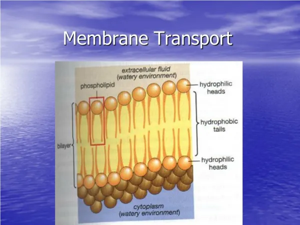
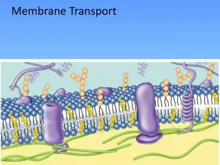
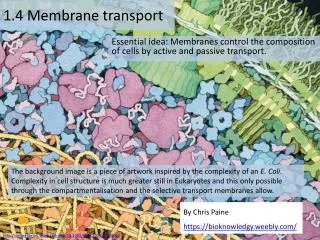
1.4 Membrane transport Essential idea: Membranes control the composition of cells by active and passive transport. The background image is a piece of artwork inspired by the complexity of an E. Coli. Complexity in cell structure is much greater still in Eukaryotes and this only possible through the compartmentalisation and the selective transport membranes allow. By Chris Paine https://bioknowledgy.weebly.com/ http://onlinelibrary.wiley.com/doi/10.1002/bmb.20345/full#fig2
1.4.U1 Particles move across membranes by simple diffusion, facilitated diffusion, osmosis and active transport.
1.4.U1 Particles move across membranes by simple diffusion, facilitated diffusion, osmosis and active transport. http://www.phschool.com/science/biology_place/labbench/lab1/concepts.html http://highered.mheducation.com/sites/0072495855/student_view0/chapter2/animation__how_diffusion_works.html
1.4.U1 Particles move across membranes by simple diffusion, facilitated diffusion, osmosis and active transport.
1.4.U1 Particles move across membranes by simple diffusion, facilitated diffusion, osmosis and active transport.
1.4.U1 Particles move across membranes by simple diffusion, facilitated diffusion, osmosis and active transport. http://www.northland.cc.mn.us/biology/Biology1111/animations/transport1.html
1.4.U1 Particles move across membranes by simple diffusion, facilitated diffusion, osmosis and active transport.
1.4.U1 Particles move across membranes by simple diffusion, facilitated diffusion, osmosis and active transport. What is osmosis? http://www.flickr.com/photos/luchilu/399970490/
1.4.U1 Particles move across membranes by simple diffusion, facilitated diffusion, osmosis and active transport. Osmosis may occur when there is a partially permeable membrane, such as a cell membrane. When a cell is submerged in water, the water molecules pass through the cell membrane from an area of low solute concentration (outside the cell) to one of high solute concentration (inside the cell) (Wikipedia) http://highered.mcgraw-hill.com/sites/0072495855/student_view0/chapter2/animation__how_osmosis_works.html Aquaporin is an integral protein that, as it’s name suggests, acts as a pore in the membrane that speeds the movement of water molecules http://opm.phar.umich.edu/protein.php?pdbid=1sor
1.4.A2 Tissues or organs to be used in medical procedures must be bathed in a solution with the same osmolarity as the cytoplasm to prevent osmosis. The importance of osmotic control http://highered.mcgraw-hill.com/sites/0072495855/student_view0/chapter2/animation__how_osmosis_works.html http://commons.wikimedia.org/wiki/File:Osmotic_pressure_on_blood_cells_diagram.svg http://commons.wikimedia.org/wiki/File:Turgor_pressure_on_plant_cells_diagram.svg
1.4.A2 Tissues or organs to be used in medical procedures must be bathed in a solution with the same osmolarity as the cytoplasm to prevent osmosis. The importance of osmotic control preventing damage to cells and tissues • Common medical procedures in which an isotonic saline solution is useful: • fluids introduction to a patient’s blood system via an intravenous drip, e.g for rehydration • used to rinse wounds, skin abrasions etc. • keep areas of damaged skin moist before applying skin grafts • eye drops/wash • frozen and used pack donor organs for transportation http://www.defenseimagery.mil/imageRetrieve.action?guid=8c9d5fade029a4f5a68fe667d1ae802ba9f30dd5&t=2
1.4.U1 Particles move across membranes by simple diffusion, facilitated diffusion, osmosis and active transport. Simple Diffusion http://commons.wikimedia.org/wiki/File:Scheme_simple_diffusion_in_cell_membrane-en.svg
1.4.U1 Particles move across membranes by simple diffusion, facilitated diffusion, osmosis and active transport. Facilitated Diffusion: Large and polar molecules can’t get across the membrane via simple diffusion Transmembrane (polytopic) proteins recognise a particular molecule and help it to move across the membrane. The direction it moves is dependent on the concentration gradient. Watch the animation http://highered.mcgraw-hill.com/sites/0072495855/student_view0/chapter2/animation__how_facilitated_diffusion_works.html http://commons.wikimedia.org/wiki/File:Scheme_facilitated_diffusion_in_cell_membrane-en.svg
1.4.A1 Structure and function of sodium–potassium pumps for active transport and potassium channels for facilitated diffusion in axons. • Potassium channels in axons are voltage gated. They enable the facilitated diffusion of potassium out of the axon • At one stage during a nerve impulse there are relatively more positive charges inside. • This voltage change causes potassium channels to open, allowing potassium ions to diffuse out of the axon. • Once the voltage conditions change the channel rapidly closes again. n.b.other positively charged ions that we might expect to pass through the pore are either too large to fit through or are too small to form bonds with the amino acids in the narrowest part of the pore - this explains the specificity of the channel. http://bioserv.fiu.edu/~walterm/human_online/nervous/nervous_system_files/image012.gif
1.4.U1 Particles move across membranes by simple diffusion, facilitated diffusion, osmosis and active transport. Primary active transport requires ATP. Integral protein pumps use the energy from the hydrolysis of ATP to move ions or large molecules across the cell membrane. Molecules are moved against their concentration gradient http://highered.mcgraw-hill.com/sites/0072495855/student_view0/chapter2/animation__how_the_sodium_potassium_pump_works.html http://commons.wikimedia.org/wiki/File:Scheme_sodium-potassium_pump-en.svg
1.4.U1 Particles move across membranes by simple diffusion, facilitated diffusion, osmosis and active transport.
1.4.U1 Particles move across membranes by simple diffusion, facilitated diffusion, osmosis and active transport.
1.4.A1 Structure and function of sodium–potassium pumps for active transport and potassium channels for facilitated diffusion in axons. The sodium–potassium pump follows a repeating cycle of steps that result in three sodium ions being pumped out of the axon (of neurons)and two potassium ions being pumped in. Each time the pump goes round this cycle it uses one ATP. The cycle consists of these steps: 1 The interior of the pump is open to the inside of the axon; three sodium ions enter the pump and attach to their binding sites. http://commons.wikimedia.org/wiki/File:Scheme_sodium-potassium_pump-en.svg
1.4.A1 Structure and function of sodium–potassium pumps for active transport and potassium channels for facilitated diffusion in axons. 3 The interior of the pump opens to the outside of the axon and the three sodium ions are released. 2 ATP transfers a phosphate group from itself to the pump; this causes the pump to change shape and the interior is then closed. http://commons.wikimedia.org/wiki/File:Scheme_sodium-potassium_pump-en.svg
1.4.A1 Structure and function of sodium–potassium pumps for active transport and potassium channels for facilitated diffusion in axons. 4 Two potassium ions from outside can then enter and attach to their binding sites. 5 6 Binding of potassium causes release of the phosphate group; this causes the pump to change shape again so that it is again only open to the inside of the axon. The interior of the pump opens to the inside of the axon and the two potassium ions are released. Sodium ions can now enter and bind to the pump again (#1). http://commons.wikimedia.org/wiki/File:Scheme_sodium-potassium_pump-en.svg
1.4.U1 Particles move across membranes by simple diffusion, facilitated diffusion, osmosis and active transport. In secondary active transport, the required energy is derived from energy stored in the form of concentration differences in a second solute. http://commons.wikimedia.org/wiki/File:Scheme_secundary_active_transport-en.svg
1.4.U1 Particles move across membranes by simple diffusion, facilitated diffusion, osmosis and active transport. Typically, the concentration gradient of the second solute was created by primary active transport, and the diffusion of the second solute across the membrane drives secondary active transport. http://bcs.whfreeman.com/thelifewire/content/chp05/0502002.html
1.4.U3 Vesicles move materials within cells. Vesicles are small spheroidal packages that bud off of the RER and the Golgi apparatus They carry proteins produced by ribosomes on the RER to the Golgi apparatus, where they are prepared for export from the cell via another vesicle Use the animated tutorial to learn more about the formation and use of vesicles in cells http://www.sumanasinc.com/webcontent/animations/content/vesiclebudding.html
1.4.U2 The fluidity of membranes allows materials to be taken into cells by endocytosis or released by exocytosis. Endocytosis: The taking in of external substances by an inward pouching of the plasma membrane, forming a vesicle Exocytosis: The release of substances from a cell (secretion) when a vesicle joins with the cell plasma membrane. http://highered.mcgraw-hill.com/olc/dl/120068/bio02.swf Diagrams on following slides
1.4.U2 The fluidity of membranes allows materials to be taken into cells by endocytosis or released by exocytosis. Constitutive secretion occurs continuously in cells, depending on their function Regulated secretion is in response to a trigger e.g. the release of neurotransmitters http://commons.wikimedia.org/wiki/File:Exocytosis_types.svg
1.4.U2 The fluidity of membranes allows materials to be taken into cells by endocytosis or released by exocytosis. “Cell drinking” “Cell eating” http://commons.wikimedia.org/wiki/File:Endocytosis_types.svg
1.4.U2 The fluidity of membranes allows materials to be taken into cells by endocytosis or released by exocytosis.
1.4.S1 Estimation of osmolarity in tissues by bathing samples in hypotonic and hypertonic solutions. (Practical 2) Estimation of osmolarityis a simple lab with many possible variations. http://www.saps.org.uk/secondary/teaching-resources/286-measuring-the-water-potential-of-a-potato-cell • The two lab protocols shown suggest different ways of measuring the dependent variable. • This is an ideal opportunity to practise and improve your understanding of the following IA criteria: • Analysis • Evaluation • Communication http://www.nuffieldfoundation.org/practical-biology/investigating-effect-concentration-blackcurrant-squash-osmosis-chipped-potatoes n.b.estimation of osmolarity should be limited to statements such as “this tissue has an osmolarity equivalent to a 2% sucrose solution”. Molar concentrations maybe used in place of % and other solution such as sodium chloride maybe used in place of glucose.
Bibliography / Acknowledgments Jason de Nys