Download
1 / 16
160 likes | 329 Views
Membrane transport. Chemistry 256. Types of ion channels (passive transport). Mechanosensitive channels – touch, sound, osmotic pressure changes Ligand-gated channels – external chemical signal (neurotransmitter) Signal-gated channels – internal chemical signal (Ca 2+ )

E N D
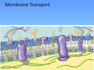
Membrane transport Chemistry 256
Types of ion channels (passive transport) • Mechanosensitive channels – touch, sound, osmotic pressure changes • Ligand-gated channels – external chemical signal (neurotransmitter) • Signal-gated channels – internal chemical signal (Ca2+) • Voltage-gated channels – membrane potential changes,
The neuron’s action potential • A nerve impulse works as a coordinated effort between a Na+ channel former and a K+ channel former. There is a high [Na+] and a low [K+] outside the cell membrane. • As the neural membrane is stimulated, the Na+ channel opens, allowing Na+ to flow into the cell, and causing nearby Na+ channels to open, which propagates the signal.
Changes in neuron membrane ion permeability • This depolarizes (reduces) the membrane potential, which causes nearby K+ channels to open, allowing K+ to flow out of the cell, which repolarizes (increases) the membrane potential. • But both gates close soon after the repolarization, and it takes a little while to bring the membrane back to a resting potential by the work of other ion pumps.
Mechanism of opening/closing a K+ channel • The S’s in the diagram below represent α-helices; S4 is the site of the “voltage sensor” and S5 and S6 are the site of the K+ channel itself. • As S4 builds up potential, the S4-S5 “linker” helix shifts, allow S6 to shift, opening the channel.
Different ions, different channel mechanisms • K+ channels work by physically opening or closing the channel (the position of the helix, the position of the N-terminus “ball”) • Cl– channels work by repulsion or lack of repulsion of the ion by a Glu.
Aquaporins • Though most cell membranes allow water to pass freely due to its small size and high concentration, some cells have such high water throughput that they need help. Aquaporins (Agre, 1992) allow high rate of water transport but are quite selective; H3O+ cannot pass through. The protein has a constriction in the center that allows one water molecule at a time to pass through and severs the “proton wire.”
Transport proteins use different conformations to perform transport • Well, we knew this from hemoglobin (RT).
Gap junctions allow fast intercellular communication • Gap junctions are a bundle of similar proteins called connexins. • The space in the center of the bundle is hollow and allows small molecules and ions to pass from cell to cell without going “outside”. • Some organs, like the heart, are topologically continuous because the cells are all connected by these junctions.
How to tell mediated (protein-assisted) from nonmediated (simple diffusion) transport • The diffusion equation is linear in concentration; that is, flux µ [solute] • So simple diffusion should yield a line in a [solute] vs. flux plot • If it doesn’t, then it is mediated transport
Transport proteins can move one or more substrate molecules • Uniport = movement of one molecule at a time (GLUT 1, for instance) • Symport = movement of two different molecules in the same direction at the same time (lactose permease carries H+ and lactose into a bacterial cell, using the H+ gradient to move lactose against its gradient (secondary active transport)) • Antiport = movement of two different molecules in opposite directions at the same time (oxalate/formate transporter)
Active transport – mostly coupled to the free energy of ATP hydrolysis to move substrates • P-type ATPase; P = phosphorylation; cation transporters • F-type ATPase; F = Fn-subunits; proton transporters • V-type ATPase; V= vacuolar; proton transporters • A-type ATPase; A = anion; anion transporters • ABC transporters; ABC = ATP-binding cassette; multiple substrate transporters
Na+-K+ ATPase (pump) antiport • For each ATP, it pumps 3 Na+ into cell and pumps 2 K+ out of cell. Critical to remove Na+ to prevent cytolysis. 70% of nerve cells’ ATP goes to this protein. • Note that a net charge separation will occur.
Two conformations of Na+-K+ pump • A particular aspartate residue can be phosphorylated only in the presence of Na+, whereas hydrolysis of the phosphorylated aspartate occurs only in the presence of K+. • Thus, two states.
Active and passive transport work in tandem • Na+-glucose transport system in intestinal epithelial cells concentrates glucose in the cell • Glucose is in low concentration in the intestinal lumen, but is driven into the cell by the Na+ gradient, which is maintained by the Na+-K+ ATPase