Download

1 / 34
340 likes | 520 Views
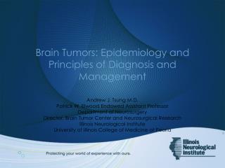
The Diagnosis and Management of Brain Abscesses Stephen Aston Stephen Aston is a Specialist Registrar in Infectious Diseases at the Tropical and Infectious Diseases Unit, Royal Liverpool University Hospital.

E N D
The Diagnosis and Management of Brain Abscesses Stephen Aston Stephen Aston is a Specialist Registrar in Infectious Diseases at the Tropical and Infectious Diseases Unit, Royal Liverpool University Hospital. Following general medical training in Birmingham, he spent time working on clinical trials in vaccine development in Oxford and the 2009-10 influenza pandemic in Liverpool, before taking up his current post. He is particularly interested in immune responses to severe bacterial infections and how this informs vaccine development. Edited by Prof Tom Solomon and Dr Agam Jung • the diagnosis and management of brain abscesses • Learning Objectives • Introduction • Development and Pathogenesis • Intracranial Infection • Pathogen Spectrum • Immunosuppressed Patients • Clinical Features • Investigation • MRI • Further Neuroimaging • Microbiological Investigations • Management • Key Points • Summary • Self Assessment This session provides a review of brain abscesses, including a brief description of aetiology and pathogenesis, later focusing on current investigation and best practice in management.
Learning Objectives • By the end of this session you will be able to: • Describe the stages of development and pathogenesis of brain abscesses • Identify the typical spectrum of pathogens implicated in the aetiology of brain abscess • Define the nonspecific nature of the clinical features of brain abscess • List the appropriate investigation of possible brain abscess and to identify the important radiological features on CT and MR scanning • Develop a management plan for patients with brain abscess, including antimicrobial treatment, surgical interventions and adjunctive therapies • the diagnosis and management of brain abscesses • Learning Objectives • Introduction • Development and Pathogenesis • Intracranial Infection • Pathogen Spectrum • Immunosuppressed Patients • Clinical Features • Investigation • MRI • Further Neuroimaging • Microbiological Investigations • Management • Key Points • Summary • Self Assessment
Introduction Brain abscess is fortunately a relatively uncommon condition, with an incidence of approximately 1 in 100,000 per year. Prior to pioneering advancements in neurosurgical techniques in the late 19th century, brain abscesses were invariably fatal. The development of effective anti-microbial agents following the Second World War secured further improvements in prognosis. With the inception of modern neuroimaging techniques allowing earlier diagnosis, mortality rates have fallen further, presently between 5 and 15%. However, debilitating sequelae such as seizures, paresis and cognitive impairment remain common. This session outlines the pathogenesis, aetiology and typical clinical features of brain abscess. Approaches to diagnosing the condition are described before reviewing current management strategies. • the diagnosis and management of brain abscesses • Learning Objectives • Introduction • Development and Pathogenesis • Intracranial Infection • Pathogen Spectrum • Immunosuppressed Patients • Clinical Features • Investigation • MRI • Further Neuroimaging • Microbiological Investigations • Management • Key Points • Summary • Self Assessment MRI (T1 + contrast) showing a small ring-enhancing lesion
Stages of Development and Pathogenesis of Brain Abscesses I Brain abscesses occur at focal points of bacterial multiplication within the brain parenchyma; they begin as a localised area of cerebritis and later progress into a collection of pus surrounded by a vascularised capsule. By virtue of the impermeability of the blood-brain barrier, the brain parenchyma is relatively resistant to the establishment of focal bacterial infection. An area of necrosis caused by for example micro-infarction or hypoxaemia is necessary to act as a nidus for bacterial multiplication. Several stages of development en route to the formation of a mature encapsulated brain abscess following bacterial ingress have been defined by neuroimaging studies • the diagnosis and management of brain abscesses • Learning Objectives • Introduction • Development and Pathogenesis • Intracranial Infection • Pathogen Spectrum • Immunosuppressed Patients • Clinical Features • Investigation • MRI • Further Neuroimaging • Microbiological Investigations • Management • Key Points • Summary • Self Assessment
Stages of Development and Pathogenesis of Brain Abscesses II • the diagnosis and management of brain abscesses • Learning Objectives • Introduction • Development and Pathogenesis • Intracranial Infection • Pathogen Spectrum • Immunosuppressed Patients • Clinical Features • Investigation • MRI • Further Neuroimaging • Microbiological Investigations • Management • Key Points • Summary • Self Assessment Early Cerebritis Days 1-3: Perivascular inflammation, characterised by neutrophil infiltration, occurs around the site of focal infection with a surrounding area of oedema. 2. Late Cerebritis Days 4-9: A central area of necrosis develops as the surrounding oedema progresses. Peripheral accumulation of fibroblasts preludes the development of a capsule. 3. Early Capsule Days 10-14: Establishment of a ring-enhancing capsule of well-vascularised tissue with further fibroblast migration and adjacent reactive astrocytosis. 4. Late Capsule Day 14 and beyond: Collagen fibre and granulation tissue deposition leads to a thickening of the capsule effectively walling off the area of focal suppurative infection.
Intracranial Infection I To establish intracranial infection, bacteria reach the brain via three main routes: Extension from a contiguous focus of infection, typically the middle ear or paranasal sinuses Haematogenous (metastatic) spread from a distant extracranial source Direct inoculation following neurosurgery or penetrating trauma However in up to 30% of cases of brain abscess, the route of infection cannot be determined. Suppurative intracranial infection can also manifest as subdural empyema, in which pus collects between the dura and arachnoid mater, and less frequently as extradural abscess, in which pus accumulates in the potential space between the inner skull table and dura mater. These conditions share a common aetiological basis with brain abscess and may occur concurrently. • the diagnosis and management of brain abscesses • Learning Objectives • Introduction • Development and Pathogenesis • Intracranial Infection • Pathogen Spectrum • Immunosuppressed Patients • Clinical Features • Investigation • MRI • Further Neuroimaging • Microbiological Investigations • Management • Key Points • Summary • Self Assessment
Intracranial Infection II • the diagnosis and management of brain abscesses • Learning Objectives • Introduction • Development and Pathogenesis • Intracranial Infection • Pathogen Spectrum • Immunosuppressed Patients • Clinical Features • Investigation • MRI • Further Neuroimaging • Microbiological Investigations • Management • Key Points • Summary • Self Assessment Brain abscess is the most common pattern of intracranial suppurative infection
Spectrum of Pathogens Implicated in the Aetiology of Brain Abscess I An understanding of the aetiology of brain abscess is important, since the primary source of infection not only correlates with the location of abscesses within the brain and hence their clinical features, but also the potential spectrum of causative organisms and thus informs the choice of antimicrobial treatment regimens. The following pages will summarise the typical spectrum of causative pathogens divided by source of infection. Initial empirical antimicrobials treatment regimens are also suggested. • the diagnosis and management of brain abscesses • Learning Objectives • Introduction • Development and Pathogenesis • Intracranial Infection • Pathogen Spectrum • Immunosuppressed Patients • Clinical Features • Investigation • MRI • Further Neuroimaging • Microbiological Investigations • Management • Key Points • Summary • Self Assessment Scanning Electron Micrograph of Staphylococcus epidermidis
Spectrum of Pathogens Implicated in the Aetiology of Brain Abscess II Otogentic and Paranasal Sinus Brain abscesses secondary to ear infections are diminishing in frequency in developed countries as a result of the widespread use of early antimicrobial treatment for suppurative otitis media. Otogenic infection remains a frequent cause of brain abscess in the developing world. Brain abscess complicating paranasal sinus infection is most commonly seen in young men. Subdural empyema is particularly associated with this route of infection. • the diagnosis and management of brain abscesses • Learning Objectives • Introduction • Development and Pathogenesis • Intracranial Infection • Pathogen Spectrum • Immunosuppressed Patients • Clinical Features • Investigation • MRI • Further Neuroimaging • Microbiological Investigations • Management • Key Points • Summary • Self Assessment
Spectrum of Pathogens Implicated in the Aetiology of Brain Abscess III Haematogenous *Additional anti-staphylococcal cover with e.g. vancomycin is recommended for presumed endocarditis-related brain abscess **Bronchiectasis, lung abscess and empyema are important causes of infection orginating in the respiratory tract Haematogenous spread is now the most common route of infection. Typically, multiple or multi-loculated abscesses are seen, occurring predominantly within the territory of the middle cerebral artery, often at the grey white matter interface where blood flow is most marginal. • the diagnosis and management of brain abscesses • Learning Objectives • Introduction • Development and Pathogenesis • Intracranial Infection • Pathogen Spectrum • Immunosuppressed Patients • Clinical Features • Investigation • MRI • Further Neuroimaging • Microbiological Investigations • Management • Key Points • Summary • Self Assessment
Spectrum of Pathogens Implicated in the Aetiology of Brain Abscess IV Haematogenous Other Sources Peridontalinfections often give to rise polymicrobial infections. Congenital heart disease complicated by a right to left shunt was a well recognised cause of brain abscess. Early diagnosis and operative repair of such lesions has seen a marked reduction in brain abscess attributable to this source. • the diagnosis and management of brain abscesses • Learning Objectives • Introduction • Development and Pathogenesis • Intracranial Infection • Pathogen Spectrum • Immunosuppressed Patients • Clinical Features • Investigation • MRI • Further Neuroimaging • Microbiological Investigations • Management • Key Points • Summary • Self Assessment
Spectrum of Pathogens Implicated in the Aetiology of Brain Abscess V Penetrating Head Trauma and Postoperative Penetrating head trauma such as gun shot wounds, open depressed skull fractures with foreign bodies in the brain parenchyma and basal skull fractures with CSF fistula can give rise to all forms of intracranial suppurative infection • the diagnosis and management of brain abscesses • Learning Objectives • Introduction • Development and Pathogenesis • Intracranial Infection • Pathogen Spectrum • Immunosuppressed Patients • Clinical Features • Investigation • MRI • Further Neuroimaging • Microbiological Investigations • Management • Key Points • Summary • Self Assessment
Brain Abscesses in Immunosuppressed Patients I • Whilst in immunocompetent individuals brain abscess are usually caused by pyogenic bacteria, in the immunosuppressed, a much broader array of organisms is implicated. • These include: • Toxoplasma gondi(shown below) • Aspergillus species • Candida species • Nocardia • Mycobacterium tuberculosis • the diagnosis and management of brain abscesses • Learning Objectives • Introduction • Development and Pathogenesis • Intracranial Infection • Pathogen Spectrum • Immunosuppressed Patients • Clinical Features • Investigation • MRI • Further Neuroimaging • Microbiological Investigations • Management • Key Points • Summary • Self Assessment This is a high power view of brain in treated cerebral toxoplasmosis. Immunocytochemical stain using a specific antibody to Toxoplasma gondii and the immunoperoxidase method – Wellcome Images.
Brain Abscesses in Immunosuppressed Patients II • Histological/Microbiological diagnosis • Moreover, in this patient group there is a high incidence of non-infective space occupying lesions such as lymphoma. • This highlights the particular importance of attempting to obtain histological and microbiological confirmation of diagnosis in these patients. • HIV • There is an important caveat to this general principle for patients with advanced HIV infection. • Here, toxoplasmosis is the likely cause of abscess-like lesions and empirical therapy should be given for two weeks. If there is no clinical or radiological improvement, diagnostic aspiration should be performed. • the diagnosis and management of brain abscesses • Learning Objectives • Introduction • Development and Pathogenesis • Intracranial Infection • Pathogen Spectrum • Immunosuppressed Patients • Clinical Features • Investigation • MRI • Further Neuroimaging • Microbiological Investigations • Management • Key Points • Summary • Self Assessment
Non-specific Nature of the Clinical Features of Brain Abscess The presentation of brain abscess is variable and relates to the location, size and number of abscesses, the virulence of the infecting microorganisms and the immunological status of the affected individual. Males are affected approximately twice as commonly as females; the median age is 30-45 years. The clinical manifestations are nonspecific, meaning that diagnosis is often delayed. They relate predominantly to a rapidly expanding intracranial mass rather than an infective process. Common presenting features are listed in the above table. • the diagnosis and management of brain abscesses • Learning Objectives • Introduction • Development and Pathogenesis • Intracranial Infection • Pathogen Spectrum • Immunosuppressed Patients • Clinical Features • Investigation • MRI • Further Neuroimaging • Microbiological Investigations • Management • Key Points • Summary • Self Assessment
Investigation I • Neuroimaging • There is no single reliable confirmatory test for the diagnosis of brain abscess. • Blood samples • Laboratory tests on blood samples are often unremarkable. • Neuroimaging • The diagnosis rests heavily on suggestive neuroimaging findings in the individuals with a compatible clinical presentation. • The availability of neuroimaging techniques permitting detailed resolution of the brain parenchyma has had a profound impact on the management and outcome of brain abscess. • The appearance of brain abscesses varies significantly with their pathological stage, hence precise clinical correlation regarding the timing of scan with respect to the onset of symptoms is required. • the diagnosis and management of brain abscesses • Learning Objectives • Introduction • Development and Pathogenesis • Intracranial Infection • Pathogen Spectrum • Immunosuppressed Patients • Clinical Features • Investigation • MRI • Further Neuroimaging • Microbiological Investigations • Management • Key Points • Summary • Self Assessment
Investigation II A mature brain abscess appears as ring-enhancing lesions, for which there is a broad differential diagnosis. • the diagnosis and management of brain abscesses • Learning Objectives • Introduction • Development and Pathogenesis • Intracranial Infection • Pathogen Spectrum • Immunosuppressed Patients • Clinical Features • Investigation • MRI • Further Neuroimaging • Microbiological Investigations • Management • Key Points • Summary • Self Assessment
MRI I • The appearances of brain abscess on MRI are similarly dependent upon their stage of evolution. • T1-weighted Images • Early cerebral abscesses appear as a poorly-defined hypointense area relative to the normal brain parenchyma. • As the abscess matures, the hypointense area becomes better demarcated and the capsule becomes apparent as a slightly hyperintense rim. • Ring-enhancement following the administration of intravenous gadolinium is usually seen. • T2-weighted Images • A mature abscess has a hyperintense central area, corresponding to the pus-filled core, and a hypointense capsule and surrounding oedema. • CT is less sensitive than MRI for the detection of brain abscess, although it has the advantages of being quick and readily performed in even the most acutely unwell patients. • The changes produced by early brain abscess on CT are subtle and may easily be overlooked on non-contrast scans. • the diagnosis and management of brain abscesses • Learning Objectives • Introduction • Development and Pathogenesis • Intracranial Infection • Pathogen Spectrum • Immunosuppressed Patients • Clinical Features • Investigation • MRI • Further Neuroimaging • Microbiological Investigations • Management • Key Points • Summary • Self Assessment
MRI II Early cerebritis appears as a focal area of low density that enhances following the administration of intravenous contrast medium. As the abscess capsule matures, the classic appearance of a ring-enhancing lesion becomes more evident • the diagnosis and management of brain abscesses • Learning Objectives • Introduction • Development and Pathogenesis • Intracranial Infection • Pathogen Spectrum • Immunosuppressed Patients • Clinical Features • Investigation • MRI • Further Neuroimaging • Microbiological Investigations • Management • Key Points • Summary • Self Assessment MRI (T1+contrast) showing a small ring-enhancing lesion
Neuroimaging – Further considerations Neuroimaging alone cannot with certainty distinguish brain abscess from other ring-enhancing lesions. Differentiating brain abscess and metastatic tumour is an important diagnostic challenge. Key features are listed below. Interval scans have some utility in this setting; a rapidly expanding lesion favours a diagnosis of cerebral abscess. Newly developed techniques such as diffusion-weighted imaging and MR spectroscopy also show some promise in differentiating abscess from necrotic neoplasms and other cystic lesions. • the diagnosis and management of brain abscesses • Learning Objectives • Introduction • Development and Pathogenesis • Intracranial Infection • Pathogen Spectrum • Immunosuppressed Patients • Clinical Features • Investigation • MRI • Further Neuroimaging • Microbiological Investigations • Management • Key Points • Summary • Self Assessment • Features suggestive of cerebral abscess: • Thin walled capsule • Thinning at the ventricular margin • Peripheral contrast enhancement • Features suggestive of metastatic tumour: • Thickened, irregular and nodular capsule • Diffuse contrast enhancement
Microbiological Investigations Choosing appropriate antimicrobial therapy for brain abscess relies on accurate identification of the causal pathogen. In all but exceptional circumstances, brain abscess material should be obtained for culture. With meticulous specimen handling, culture yields approach 100% for samples obtained prior to starting antimicrobials. Culture yields fall dramatically for specimens obtained after treatment has started. The Gram stain may, however, remain positive which alone may provide a useful guide to management. When obtained, specimens should be cultured for aerobic and anaerobic bacteria, mycobacteria and fungi. Other clues to the likely causative pathogen may be obtained by culture of material from sites presumed to be the primary focus of infection (e.g. paranasal sinus). CSF analysis, however, rarely provides any useful information and does not justify the substantial risk of brainstem herniation that lumbar puncture entails. • the diagnosis and management of brain abscesses • Learning Objectives • Introduction • Development and Pathogenesis • Intracranial Infection • Pathogen Spectrum • Immunosuppressed Patients • Clinical Features • Investigation • MRI • Further Neuroimaging • Microbiological Investigations • Management • Key Points • Summary • Self Assessment
Brain Abscess Management I • Overview • There are limited data from good quality clinical trials to guide the management of brain abscess and historically clinical practices have been diverse. • The main treatment strategies are: • Antimicrobial agents • Needle aspiration - often using stereotactic guidance • Complete surgical excision • In addition, adjunctive therapies such as corticosteroids and anticonvulsant agents are variably used. • A combination of antimicrobial therapy and aspiration is now used for the majority of cases, with medical therapy alone and complete surgical excision reserved for particular circumstances. • the diagnosis and management of brain abscesses • Learning Objectives • Introduction • Development and Pathogenesis • Intracranial Infection • Pathogen Spectrum • Immunosuppressed Patients • Clinical Features • Investigation • MRI • Further Neuroimaging • Microbiological Investigations • Management • Key Points • Summary • Self Assessment
Brain Abscess Management II . • the diagnosis and management of brain abscesses • Learning Objectives • Introduction • Development and Pathogenesis • Intracranial Infection • Pathogen Spectrum • Immunosuppressed Patients • Clinical Features • Investigation • MRI • Further Neuroimaging • Microbiological Investigations • Management • Key Points • Summary • Self Assessment
Brain Abscess Management III • Antimicrobial therapy • Effective antimicrobial therapy for brain abscess relies on identification of the causative microorganism allowing the selection of an appropriate regimen. Ideally, it therefore follows an aspiration procedure. • There are several important factors to consider in choosing an appropriate treatment regimen. • Activity against implicated pathogens • Before culture information is available, the probable causative bacteria are predicted on the basis of the putative source of infection and the location of the abscess. • Ability to penetrate abscess cavity • There is limited data available to guide practice. • Inferences are made from studies of CSF antimicrobial concentrations in the treatment of bacteria meningitis, although the characteristics of the blood-brain and blood-CSF barrier are known to differ. • . • the diagnosis and management of brain abscesses • Learning Objectives • Introduction • Development and Pathogenesis • Intracranial Infection • Pathogen Spectrum • Immunosuppressed Patients • Clinical Features • Investigation • MRI • Further Neuroimaging • Microbiological Investigations • Management • Key Points • Summary • Self Assessment
Brain Abscess Management IV • Duration and route of treatment • Lone medical management • Medical management alone is sometimes used in patients who are regarded as poor candidates for surgery. It is preferable if the causative pathogen can be inferred from results of positive cultures from primary sites of infection, otherwise an empirical regimen is selected. • It may also be considered for… • Multiple small abscesses • Abscesses located in surgically unaccessible or eloquent areas • It is most likely to be successful if… • Abscesses are small (i.e. <1.5cm) • In cerebritis stage • Located in a well vascularised cortical area • Frequent interval scans should be performed to assess therapeutic response and to identify complications requiring definitive surgical management. • the diagnosis and management of brain abscesses • Learning Objectives • Introduction • Development and Pathogenesis • Intracranial Infection • Pathogen Spectrum • Immunosuppressed Patients • Clinical Features • Investigation • MRI • Further Neuroimaging • Microbiological Investigations • Management • Key Points • Summary • Self Assessment
Brain Abscess Management V • Duration and route of treatment • Corticosteroids • Adjuvant corticosteroids are often used to reduce vasogenic oedema associated with brain abscesses. • There are important concerns regarding steroid use… • Effectiveness in reducing oedema and mass effect not established in human clinical trials • May retard abscess capsule development • May reduce antimicrobial penetration • Give false impression of a therapeutic response by reducing ring-enhancement on follow-up scans • Most authors recommend that corticosteroids are reserved for situations of raised intracranial pressure resulting in a clear risk of brainstem herniation or other significant neurological deficit. • the diagnosis and management of brain abscesses • Learning Objectives • Introduction • Development and Pathogenesis • Intracranial Infection • Pathogen Spectrum • Immunosuppressed Patients • Clinical Features • Investigation • MRI • Further Neuroimaging • Microbiological Investigations • Management • Key Points • Summary • Self Assessment
Brain Abscess Management VI • Duration and route of treatment • Anticonvulsants • Seizures are a frequent complication of brain abscess both in the acute setting and for a prolonged period after the resolution of the acute infection. • Some advocate the use of seizure prophylaxis for extended periods in every case of brain abscess, although there are again few good data. • If commenced, anticonvulsants should probably be continued for 6-12 months and then only withdrawn if the patient is seizure-free, the EEG normal and no signs of on going inflammation on neuroimaging. • the diagnosis and management of brain abscesses • Learning Objectives • Introduction • Development and Pathogenesis • Intracranial Infection • Pathogen Spectrum • Immunosuppressed Patients • Clinical Features • Investigation • MRI • Further Neuroimaging • Microbiological Investigations • Management • Key Points • Summary • Self Assessment
Brain Abscess Management VII • Surgical • Surgical techniques for the management of brain abscess have evolved significantly in recent years. • Whilst debate still continues as to the best approach in particular situations, minimally-invasive aspiration and closed-drainage procedures have largely replaced craniotomy and attempted excision as the first line strategy. • Aspiration is not considered appropriate in all cases of brain abscess. Circumstances when excision may be the preferred initial surgical option are listed in the table on the next slide. • The most effective strategy for abscesses that do not improve or recur following attempted aspiration is not established. Repeated aspiration may be attempted, sometimes with the placement of a catheter to allow on going drainage and instillation of intracavityantimicrobials. • Alternatively complete surgical excision is undertaken. • the diagnosis and management of brain abscesses • Learning Objectives • Introduction • Development and Pathogenesis • Intracranial Infection • Pathogen Spectrum • Immunosuppressed Patients • Clinical Features • Investigation • MRI • Further Neuroimaging • Microbiological Investigations • Management • Key Points • Summary • Self Assessment
Brain Abscess Management VIII • the diagnosis and management of brain abscesses • Learning Objectives • Introduction • Development and Pathogenesis • Intracranial Infection • Pathogen Spectrum • Immunosuppressed Patients • Clinical Features • Investigation • MRI • Further Neuroimaging • Microbiological Investigations • Management • Key Points • Summary • Self Assessment
Brain Abscess Management IX • Adjunctive therapies • As described in the introduction, advanced neurosurgical practices and potent antimicrobial agents, supported by modern neuroimaging technologies, have reduced mortality from brain abscess from being nearly universal to current levels of around 10%. • However, significantly decreased level of consciousness at presentation remains a poor prognostic indicator. In one series, 62% of patients with a GCS of <9 either died or fell into a permanent vegetative state. • Major sequelae such as seizures, focal neurological deficit and cognitive impairment are common in survivors. • Seizures occur in up to 50% of patients and may have a latent period as long as 5 years. • Focal neurological deficit occur in 25-50%. • Significant cognitive impairment occurs in 20% of survivors, although is probably underreported. • Recurrence rates of between 10-50% have been reported. Surveillance is recommended for at least 1 year • the diagnosis and management of brain abscesses • Learning Objectives • Introduction • Development and Pathogenesis • Intracranial Infection • Pathogen Spectrum • Immunosuppressed Patients • Clinical Features • Investigation • MRI • Further Neuroimaging • Microbiological Investigations • Management • Key Points • Summary • Self Assessment
Key Points • Brain abscesses are relatively uncommon and mortality is now around 10%. However, significant attributable morbidity still occurs in affected individuals. • Focal multiplication of bacteria at sites of parenchymal damage (often microscopic) lead to the formation of brain abscess. • Bacteria reach the brain parenchyma via spread from a contiguous skull-vault infection, haematogenous spread from a distant site or direct inoculation following trauma or surgery. • In immunocompetent individuals, abscesses are caused by a broad but often predictable array of bacterial pathogens. • The clinical features are nonspecific and relate mainly to an expanding intracranial mass rather than an infective process. • Modern neuroimaging techniques have dramatically improved our ability to detect brain abscesses, but microbiological confirmation is vital for directing treatment. • Most brain abscesses can be managed with a combination of surgical aspiration followed by appropriate antimicrobial therapy • the diagnosis and management of brain abscesses • Learning Objectives • Introduction • Development and Pathogenesis • Intracranial Infection • Pathogen Spectrum • Immunosuppressed Patients • Clinical Features • Investigation • MRI • Further Neuroimaging • Microbiological Investigations • Management • Key Points • Summary • Self Assessment
Summary • Having completed this session you will now be able to: • Describe the stages of development and pathogenesis of brain abscesses • Identify the typical spectrum of pathogens implicated in the aetiology of brain abscess • Define the nonspecific nature of the clinical features of brain abscess • List the appropriate investigation of possible brain abscess and to identify the important radiological features on CT and MR scanning • Develop a management plan for patients with brain abscess, including antimicrobial treatment, surgical interventions and adjunctive therapies • References • Mathisen GE, Johnson JP. Brain abscess. Clin Infect Dis 1997;25:763-81. • Solomon T. Intracranial space occupying infections and neurological HIV disease. In: Donaghy M (Eds). Brain's Diseases of the Nervous System - 12th Edition. Oxford University Press, USA; 2009. • Lu C-H, Chang W-N, Lui C-C. Strategies for the management of bacterial abscess. J ClinNeurosci 2006;13:979-985. • Erdogan E, Cansever T. Pyogenic brain abscess. Neurosurg Focus 2008;24:1-10. • the diagnosis and management of brain abscesses • Learning Objectives • Introduction • Development and Pathogenesis • Intracranial Infection • Pathogen Spectrum • Immunosuppressed Patients • Clinical Features • Investigation • MRI • Further Neuroimaging • Microbiological Investigations • Management • Key Points • Summary • Self Assessment
Self Assessment Question 1 • the diagnosis and management of brain abscesses • Learning Objectives • Introduction • Development and Pathogenesis • Intracranial Infection • Pathogen Spectrum • Immunosuppressed Patients • Clinical Features • Investigation • MRI • Further Neuroimaging • Microbiological Investigations • Management • Key Points • Summary • Self Assessment After how many days following initial bacterial ingress does ring-enhancement typically first become evident on neuroimaging studies? Less than 3 days 4-7 days 7-11 days 11-14 days More than 14 days
To learn more about neurological infectious diseases… NeuroID 2013: Liverpool Neurological Infectious Diseases Course Liverpool Medical Institution, UK Provisional date: May 2013 Ever struggled with a patient with meningitis or encephalitis, and not known quite what to do? Then the Liverpool Neurological infectious Diseases Course is for you! For Trainees and Consultants in Adult and Paediatric Neurology, Infectious Diseases, Acute Medicine, Emergency Medicine and Medical Microbiology who want to update their knowledge, and improve their skills. • Presented by Leaders in the Field • Commonly Encountered Clinical Problems • Practical Management Approaches • Rarities for Reference • Interactive Case Presentations • State of the Art Updates • Pitfalls to Avoid • Controversies in Neurological Infections Feedback from previous course: “Would unreservedly recommend to others” “An excellent 2 days!! The best course for a long time” Convenors: Prof Tom Solomon, Dr EnitanCarrol, Dr Rachel Kneen, Dr Nick Beeching, Dr Benedict Michael For more information and to REGISTER NOW VISIT: www.liv.ac.uk/neuroidcourse