Download

1 / 24
240 likes | 384 Views
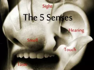
Special Senses. Taste, Smell, Sight, Hearing (we’re just going to focus on “Sight & Hearing”). Vision. Photoreceptors – visual receptor cells Adult eye = ~ 1 in. diameter Accessory structures – protect the eye or aid in its functioning Eyebrows Eyelids Conjunctiva

E N D
Special Senses Taste, Smell, Sight, Hearing (we’re just going to focus on “Sight & Hearing”)
Vision • Photoreceptors – visual receptor cells • Adult eye = ~ 1 in. diameter • Accessory structures – protect the eye or aid in its functioning • Eyebrows • Eyelids • Conjunctiva • Lacrimal apparatus • Extrinsic Eye muscles
Eyebrows & Eyelids • Eyebrows • Shade eyes from sunlight • Prevent perspiration from entering eyes • Eyelids (palpabrae) • Blinking occurs every 3-7 secs to prevent dehydration of eyes • Eyelashes are richly innervated, so anything that touches them, including a puff of air, triggers reflex blinking
Conjunctiva • Transparent mucous membrane that lines eyelids & reflects over surface of eyeball • Lubricate eye & to prevent invasion to posterior portion of eye • Conjunctivitis is an inflammation of the conjunctiva • Pinkeye is a type of conjunctivitis caused by bacteria or virus
Lacrimal Apparatus • Consists of the lacrimal gland & lacrimal ducts • Lacrimal gland releases fluid that is spread over eye when blinking • Contains mucus, antibodies & lysozyme (a bacteria-destroying enzyme)
Label your wkst! Don’t need to know this! Don’t need to know this!
Structure of the Eyeball • Made up of three layers called tunics • Fibrous (1) • Vascular (2) • Sensory (3) • Fibrous tunicis the outermost coat of the eye (1) • Divided into 2 major regions: sclera & cornea • Sclera (tough connective tissue) - “whites of the eye” • Functions to protect & shape eyeball • Sturdy anchoring for extrinsic eye muscles 3
More on the Eye • Cornea - anterior 6th of fibrous tunic • Covered on both sides by simple squamous epithelium • Lined with pain fibers (which is why contacts can be so tough to adjust to) • When cornea is touched, reflex blinking & increased lacrimal fluid secretion occur • FUN FACT: • Since cornea has no blood supply it is the only tissue that can be transplanted with very little fear of rejection (does not have contact with immune system)
Iris • Most anterior part of vascular tunic(middle layer) (2) • Between cornea & lens • Round central opening (pupil) allows light to enter eye • Made of smooth muscle fibers that contract & dilate depending on light stimulus 3
Iris • Though it seems to appear in many colors (Iris means “rainbow”), it actually only contains brown pigment • When an iris contains a lot of pigment, the eyes appear brown or black • If the amount of pigment is small, the short wavelengths of light are scattered from the unpigmented parts of the iris & eyes appear blue, green, or gray • Why, then, do newborn babies often appear to have gray or blue eyes?
The Sensory Tunic (Retina & Lens) • Deepest layer • Contains the lens(hard disc) which allows an image that is upside down & backwards • Has pigmented cells that absorb light • Stores Vitamin A, which is needed by photoreceptor cells • Contains millions of photoreceptors • Rods & cones • Rods - more numerous & are our dim-light & peripheral receptors (more sensitive to light) • Cones - bright light & provide high-acuity color vision • The optic disc (located where the optic nerve leaves the posterior portion of the eye) is called the “blind spot” because it contains no photoreceptors Lens
Internal Chambers • Filled with aqueous humorwhich is produced in posterior chamber & drains from anterior chamber • If drainage is blocked, pressure within eye may increase & cause compression of retina and optic nerve condition called glaucoma • Exam to diagnose is simple…a puff of air at the sclera will produce a measurable amount of deformation
The Ear • Divided into 3 major regions: • Inner ear • Middle ear • Outer ear
LABEL & COLOR-CODE YOUR WKST Outer EarMiddle EarInner Ear
Outer Ear • Consists of the auricle & the external auditory canal • Auricle • helix (rigid portion) • lobule (no cartilage) • Functions to direct sound waves into external auditory canal • External auditory canal • Short (~2.5 cm) & curved • Extends to the tympanic membrane (“eardrum”)
Middle Ear • Small, air-filled cavity within the temporal bone • Eustachian tubelinks middle ear to superior-most part of the throat • Normally this is closed, but yawning & swallowingopens this tube briefly to equalize pressure
Middle Ear • Contains the 3 smallest bones in the body: the ossicles • Malleus – secured to the tympanic membrane • Incus • Stapes – connects to the inner ear (via the oval window) • Tensor tympanimuscle attaches auditory tube (Eustachian tube) to malleus • This muscle helps prevent damage to inner ear under extremely loud conditions
Inner Ear • Located deep within the temporal bone & posterior to the eye socket • Made up of the vestibule, semicircular canals &cochlea
Vestibule • Central egg-shaped cavity that medially borders the middle ear • Contains perilymph (similar to CSF) • Houses equilibrium censors called maculae that respond to pull of gravity & report changes of head position
Semicircular Canals • Made up of an anterior, posterior, & lateral canals • Have receptors to help with equilibrium
Cochlea • About 1/2 size of a pea • Contains 3 hollow cavities • Cochlear duct contains spiral organ of Corti – receptor organ for hearing • Cochlear nerveruns from the spiral organ of Corti to the brain
Hair Cells in the Spiral Organ of Corti • Roughly 16,000 hearing receptor cells called cochlear hair cells line the spiral organ of Corti
Hearing…Hear’s How it Works • Sounds set up vibrations in air that beat against the ear drum • This pushes the ossicles that press fluid in the inner ear against membranes • This pressure on the membranes pulls on tiny hair cells that stimulate nearby neurons that give rise to impulses that travels to the brain, where they are interpreted
Deafness • 2 types: • Conduction • Sensorineural • Conduction deafness – occurs when something interferes with conduction of sound vibrations to the fluids of the inner ear • Sensorineural deafness – results from damage to neural structures at any point in the hearing pathway • This typically results from the gradual loss of hearing receptor cells: • Throughout life • Single explosively loud noise • Prolonged exposure to high-intensity sounds, which cause these cells to stiffen Outer & Middle Ear Inner Ear & Sensory Neurons to Brain http://www.nbclearn.com/portal/site/learn/science-of-nhl-hockey