Download
1 / 27
320 likes | 971 Views
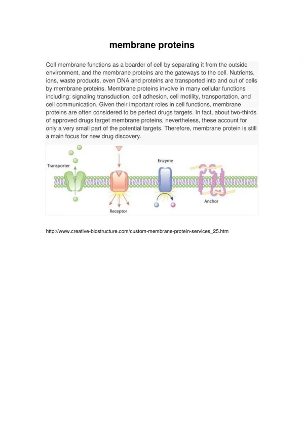
Refolding of membrane proteins for structural studies. Lars Linden * RAMC 2005. Membrane proteins as drug targets. 25% of the human genes encode for membrane proteins. The human genome:. 25%. 75%. 67% of the known drug targets are membrane proteins. 67%. 33%. The known drug targets:.

E N D
Refolding of membrane proteins for structural studies Lars Linden * RAMC 2005
Membrane proteins as drug targets • 25% of the human genes encode for membrane proteins The human genome: 25% 75% • 67% of the known drug targets are membrane proteins 67% 33% The known drug targets: membrane proteins soluble proteins m-phasys is the only company focussed exclusively on 3D structures of membrane protein targets
No human GPCR structure solved All known protein structures:~ 28,000 PDB Membrane protein structures: ~ 100 (mostly bacterial proteins) GPCR structures: 1 (Rhodopsin from bovine retina) Human GPCR structures: 0 Why ?
DNA Expression system Purification & Crystallization 3D structure Expression in Inclu-sion bodies crystal Detergent Solubilized protein Refolded protein Barriers in membrane protein structural analysis... ... and how to get around them
E. coli ? Fast Cheap High yields Multiple strains available Multiple plasmids available Selenomethionine derivatives Less time for expression = more time for crystallization! In 2004, 67% of all structures deposited in the PDB were from proteins expressed in E. coli
Expression of membrane proteins in E. coli can be toxic • Eukaryotic membrane proteins are not readily inserted into bacterial membranes • Bacterial insertion machinery becomes jammed • Protein production stops after 1 min • Low yields Possible solution: Prevent membrane insertion
Does in vitro refolding of membrane proteins work ? • Critical issues: • Energy landscape in micelles? • Non-vectorial insertion • Local vs. Global minimum?
Does in vitro refolding of membrane proteins work ? • Yes! • Bacteriorhodopsin • Light harvesting complex LHC2 • Mitochondrial transporters • Diacyl glycerol kinase • Olfactory receptor OR5 • Potassium channel KcsA • DsbB • Leukotriene receptor BLT1
Expression vector for GST-GPCR-(His)6 fusions HisTag GPCR rrBT1T2 Protease cleavage site GST AP r pGEX2a-GPCR-His Ptac ~6000 bp ori • Expression in E.coli • Preparation of inclusion bodies • Typical yields: 2-50 mg / l lac I
How to identify refolding conditions Inclusion Bodies (Aggregated Protein) Solubilisation Solubilised, but misfolded protein Detergent exchange Misfolded Refolded & native Re-aggregated
Purification and quality control of GPCRs GPCRs are rigorously testedfor activity and homogeneitybefore crystallization *) proprietary functional assay applicableto all GPCRs (including orphans)
Arrestin activity assay • Arrestin mutant binds to GPCRs constitutively • Doesn't require phosphorylation • Affinity depends on ligand binding • Requires folded GPCR 1. Bind & wash 2. Detect bound arrestin R R A A A • GPCR properly folded A R R A • GPCR not properly folded
Refolded GPCRs are functionalExample: CXCR1 Ligand binding G protein activation Bound GTPgS [dpm] Bound ligand [dpm] Log Interleukin-8 [M] Log Interleukin-8 [M] • Conclusion: • Ligand affinity (KD) like native receptor • > 80% refolded (Bmax) • Conclusion: • Couples to Gi/o • EC50 like native receptor Refolded GPCR binds ligand and couples to G protein
Refolded GPCRs are homogenousExample: CXCR1 SDS-PAGE SEC 1 2 3 - GPCR dimer - GST-GPCR fusion - GPCR monomer Absorption Volume [ml] • Inclusion body fraction • Ni chelate purified • SEC purified Superdex 200 • Conclusion: • 95 % pure on SDS gel • Conclusion: • 85 % pure by SEC analysis Refolded CXCR1 is >90% pure and monodisperse
Refolded GPCRs are homogenousExample: GPR3 Absorption Volume [ml]
Refolded GPCRs form crystals • Crystallized • Pipeline Rhodopsin family b g a d
Optimization of crystallization conditions: strategy • Truncated mutants (N- and C-termini, long loops) • Co-crystallization with ligands (agonists, antagonists, inverse agonists) • Co-crystallization with binding proteins (ß-arrestin, G proteins, antibody fragments) • Stabilization with lipids • Variation of crystallization method: vapour diffusion, microbatch, lipidic cubic phases, free interface diffusion • Selection for more thermostable mutants
Anti-GPCR monoclonal antibodies • Successful programs with antibody companies and academic groups • Refolded GPCRs used as immunogen or panning target • Antibodies obtained from mice (IgG) and phage display systems (scFvs and Fabs) • Antibodies recognize native GPCRs (FACS) • Affinity from 1 nM to 1 µM • Some are antagonistic • Some have conformation-specific epitopes Apart from their use in co-crystallization, antibodies might be used as diagnostic tools or therapeutics
m-fold CXCR1-antibody complex formation A: CXCR1-receptor B: anti-CXCR1 mAB 9D1 C: co-complex BSA mAU mAU mAU CXCR1 9D1 CXCR1 + 9D1 Aggregate 100 60 30.0 80 40 20.0 60 40 10.0 20 20 0 0.0 0 ml 8.0 12.0 14.0 ml 6.0 10.0 ml 12.0 6.0 8.0 10.0 14.0 6.0 10.0 8.0 12.0 14.0 • Immunization with CXCR1 Liposomes • Monoclonal IgG, FACS and ELISA positiv • Ligand (IL-8) is displaced by antibody (IC50 = 0,33 nM) • CXCR1 receptor and 9D1 antibody form a stable complex • scFv cloned, expressed and purified -> Co-crystallisation
Bacterial and human ion channels • Potassium Channels : voltage gated KvLQT4 hERG Kv1.3 VIC (Salmonella t.) MJKch (Methanococcus j.) Ca2+ activated KCa4 • Cloning and expression of different constructs of hERG, Kv1.3, KCa4 transmembrane region S1-S6
Bacterial and human ion channels • Ion channels are easily purified • Refoldung screen for hERG, Kv1.3, KCa4, VIC and MJKch • Tetramerisation can be detected on modified SDS or blue native Gels VIC hERG 116 tetramer 116 66 66 45 45 tetramer 35 35 132 monomer 25 25 66 18 18
Potassium channel can be produced with M-FOLD™ 116 66 45 35 25 18 • Conclusion: • Refolded K channel forms tetramer • > 95 % refolded 116 66 refolded unfolded 45 35 25 18 Refolded K channels reconstituted into planar bilayer (BLM) Refolding works for K channels
Ion channel crystals diffract to 12 Å K channel crystals