Download
1 / 3
30 likes | 64 Views
A 30-year-old man is admitted to the hospital for severe constant abdominal pain with nausea and vomiting since the previous day.

E N D
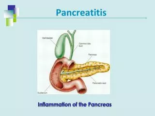
Pancreatitis Case File https://medical-phd.blogspot.com/2021/03/pancreatitis-case-file.html Eugene C. Toy, MD, Lawrence M. Ross, MD, PhD, Han Zhang, MD, Cristo Papasakelariou, MD, FACOG CASE 22 A 30-year-old man is admitted to the hospital for severe constant abdominal pain with nausea and vomiting since the previous day. He states that the pain radiates straight to his back and feels “like it’s boring a hole right through me from front to back.” He reports no other medical problems, but drinks one or two 6-packs of beer each weekend. He denies having diarrhea or fever. The serum amylase and lipase levels are markedly elevated. ⯈ What is the most likely diagnosis? ⯈ What is the anatomical location of the structure involved? ANSWER TO CASE 22: Pancreatitis Summary: A 30-year-old man who drinks alcohol is admitted to the hospital for severe abdominal pain with nausea and vomiting for 24-h duration. He states that the pain radiates straight to his back. The serum amylase and lipase levels are markedly elevated. • Most likely diagnosis: Acute pancreatitis • Anatomical location of the structure affected: Retroperitoneal, posterior to the stomach and the lesser peritoneal sac (omental bursa) CLINICAL CORRELATION The pancreas is a retroperitoneal organ, posterior to the stomach and lesser sac, partly surrounded by the duodenum. It is an exocrine gland that secretes digestive enzymes and an endocrine gland that produces insulin and glucagon to regulate blood glucose levels. Noninfectious inflammation of the pancreas is most commonly caused by alcohol abuse or gallstones. The inflammation is secondary to autodigestion of the pancreatic tissue by the exocrine secretions. Marked vomiting is typical, and serum amylase or lipase levels are elevated. Immediate management includes restricting oral intake, monitoring fluid and electrolyte balance, and pain control. The pancreatitis sometimes may be so severe as to produce hemorrhage into the pancreas or pulmonary injury. These complications are associated with higher mortality rates. APPROACH TO: The Pancreas Objectives
1. Be able to describe the anatomy of the pancreas and its relations to the duodenum and spleen 2. Be able to describe the retroperitoneal relations of the pancreas DEFINITIONS PANCREATITIS: Inflammation of the pancreas RETROPERITONEAL: Posterior or external to the peritoneal cavity OMENTAL BURSA: Subdivision of the peritoneal cavity posterior to the stomach and lesser omentum DISCUSSION The pancreas is a retroperitoneal gland that is exocrine (secretes digestive enzymes released into the duodenum) and endocrine (source of insulin and glucagon released into the bloodstream). It lies posterior to the omental bursa (lesser sac). The gland is anatomically divided into head, neck, body, and tail regions and is diagonally placed across the posterior abdominal wall (Figure 22-1). The head of the pancreas lies within the curve of the second and third parts of the duodenum, and its inferior portion forms a hooklike uncinate process that lies posterior to the superior mesenteric vessels. The neck lies at the L1 vertebral level, with the pylorus of the stomach immediately superior. The portal vein is formed posteriorly by the union of the splenic vein and superior mesenteric vein (SMV). The body of the gland passes superiorly to the left, with the tortuous splenic artery along its superior border. The short tail of the pancreas lies within the splenorenal ligament and may contact the hilum of the spleen (Table 22-1). The exocrine pancreas is drained by a main pancreatic duct, which begins in the tail and passes to the right through the body, neck, and inferior portion of the head. The duct pierces the wall of the second part of the duodenum in close association with the common bile duct, with which it typically unites to form the hepatopancreatic ampulla, which, in turn, opens through the major duodenal papilla. Several smooth muscle sphincters surround these ducts, which may enter the duodenum separately at the papilla. The superior portion of the head is drained by an accessory pancreatic duct that usually joins the main duct but may drain separately into the duodenum at the minor duodenal papilla. The head of the pancreas receives
Figure 22-1. The pancreas and its blood supply. (Reproduced, with permission, from Lindner HH. Clinical Anatomy. East Norwalk, CT: Appleton & Lange, 1989:346.)