Pancreatitis
Pancreatitis. Ultrasound Rotation Presentation Missy Purcell 3/5/10. The Pancreas a brief overview…. The pancreas has both endocrine and exocrine functions Exocrine pancreas is made up of pancreatic acinar cells and a duct system that opens into the proximal duodenum

Pancreatitis
E N D
Presentation Transcript
Pancreatitis Ultrasound Rotation Presentation Missy Purcell 3/5/10
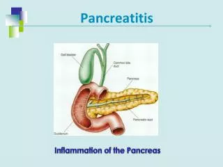
The Pancreasa brief overview… • The pancreas has both endocrine and exocrine functions • Exocrine pancreas is made up of pancreatic acinar cells and a duct system that opens into the proximal duodenum • Acinar cells synthesize and secrete digestive enzymes that are essential to digestion • The exocrine pancreas also secretes large amounts of bicarbonate, which buffers gastric acid
Pancreatitis • Pancreatitis is the most common exocrine pancreatic disease in both dogs and cats • Acute or chronic depending on whether or not the disease has lead to permanent changes in the pancreatic parenchyma • Either one can be severe and associated with pancreatic necrosis and systemic complications • Not always important to distinguish between the two
Pancreatitis • Many causes but most cases are idiopathic • Many different insults lead to pancreatitis by the same mechanism • Secretion of pancreatic juice initially decreases • This is followed by localization of zymogen granules and lysosomes, leading to the activation of tripsinogen • Tripsinogen activates more tripsinogen and other zymogens
Pancreatitismechanism continued… • Prematurely activated enzymes lead to local damage of the exocrine pancreas with pancreatic edema, bleeding, inflammation, necrosis, and peripancreatic fat necrosis • Inflammatory process leads to the recruitment of WBC and cytokine production • Activated enzymes and cytokines circulate in the bloodstream and lead to distant complications • Inflammation, DIC, multiorgan involvement or even failure
Pancreatitisclinical signs • Most common clinical signs in dogs include • Vomiting, diarrhea, weakness, abdominal pain Most common clinical signs in cats include lethargy, anorexia, dehydration, hypothermia, and abdominal pain
PancreatitisWhat to look for • In dogs a history of dietary indiscretion combined with vomiting and abdominal pain is suggestive, but most cats present with nonspecific histories and clinical sings • CBC, CHEM may suggest inflammatory changes, or show non specific changes in enzymes
PancreatitisDiagnostics • Imaging! ( you knew I would get to it sooner or later !) • Radiographic changes • Ultrasound changes
PancreatitisImaging • Radiographic findings • Both acute and chronic pancreatitis, as well as pancreatic neoplasia can result in changes visible on survey abdominal radiographs. Potential radiographic signs include: 1. Loss of abdominal detail, primarily in the right cranial abdomen, due to focal peritonitis 2. Mass effect in the right cranial abdomen 3. Displacement of the pylorus cranially, or to the left 4. Ventral or right sided displacement of the descending duodenum 5. Caudal displacement of the transverse colon 6. Bowel loops adjacent to the pancreas (usually duodenum) may be gas-filled (ileus), or corrugated/spastic in appearance
PancreatitisRadiographs • Radiographs may also be normal in dogs and cats with clinical signs of pancreatitis • Diagnosis based on radiographs is unreliable
PancreatitisUltrasound • Abdominal ultrasound has become the imaging modality of choice to help diagnose pancreatitis • Abdominal ultrasound may be completely normal in animals with pancreatitis • Changes in ultrasound images may include: • Pancreatic enlargement and fluid accumulation around the pancreas, changes in echogenicity (hyper incases of fibrosis, hypo incases of necrosis), pancreatic mass effect, hyperechogenicity of surrounding fat, dilated panceatic duct