Receptors for Perception: External and Internal Sensations
Explore how receptors like exteroceptors (vision, hearing, smell, touch), interoceptors (internal organs), and proprioceptors (body position) function. Learn about sensory neurons, muscle spindles, Golgi tendon organs, and more.

Receptors for Perception: External and Internal Sensations
E N D
Presentation Transcript
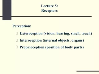
Lecture 5: Receptors Perception: • Exteroception (vision, hearing, smell, touch) • Interoception (internal objects, organs) • Proprioception (position of body parts)
Spinal cord Ganglion Neuron body T-shaped axon Sensory ending Proprioceptor Neuron The body of a sensory neuron is located in a ganglion near the spinal cord. One branch of its T-shaped axon goes to the peripheral sensory ending, and another branch goes through the dorsal roots into the spinal cord.
Proprioceptor Neuron • Body, long T-shaped axon, and sensory ending • Body is in spinal ganglia • Sensory endings generate APs in response to a specific stimulus (deformation, light, etc.) • Antidromic conduction • No dendrites, no synapses on the body
Extrafusal fiber Ia II BFdyn BFst CF Extrafusal fiber Muscle Spindle A muscle spindle is oriented parallel to extrafusal muscle fibers. It is covered with a capsule and contains two types of intrafusal muscle fibers: bag fibers (BF) and chain fibers (CF). Two types of sensory endings can be found in muscle spindles: primary (Ia) and secondary (II). Primary endings are typically seen in virtually all intrafusal fibers. Secondary endings are seen in CF and in static BF, but not in dynamic BF.
Length Action potentials Time Spindle Response to Stretch (Primary Ending) Typical responses of a primary spindle ending to an externally imposed muscle stretch at different velocities. The response increases with muscle length and with the velocity of the stretch.
Length Action potentials Time Spindle Response to Stretch (Secondary Ending) A typical response of a secondary spindle ending to an externally imposed muscle stretch and shortening. The response increases with muscle length and does not depend on velocity.
g -motoneuron dyn g -motoneurons st BFdyn BFst CF Gamma Motoneurons There are two types of small motoneurons (g-motoneurons) innervating intrafusal fibers of muscle spindles. • Dynamic g-motoneurons innervate dynamic bag fibers and change the sensitivity of primary endings. • Static g-motoneurons innervate static bag and chain fibers. They change the sensitivity of primary and secondary endings.
Length g-dynamic stimulation Time Gamma Motoneurons: Effects on Spindle Reaction to Stretch The effects of an activity of dynamic g-motoneurons on a response of a primary spindle ending to muscle stretch and shortening. In the lower graph, a g-dynamic stimulation was applied during the same changes in muscle length.
Muscle Spindle • Two types of endings: • Primary (Ia afferents): sensitive to length and velocity of muscle fibers • Secondary (II afferents): sensitive only to length of muscle fibers • Gamma-motoneurons: • A system to modify sensitivity of the spindle endings • Gamma-MNs innervate intrafusal muscle fibers
Ganglion Ib afferent Golgi tendon organ Muscle fibers Tendon Golgi Tendon Organs Golgi tendon organs are located in series with extrafusal muscle fibers at their junction with the tendon. They are innervated with fast-conducting Ib axons of sensory neurons in spinal ganglia.
Muscle force Action potentials Time Golgi Tendon Organs: Response to Muscle Force A response of a Golgi tendon organ to muscle force. Note that it is similar to the response of secondary spindle endings to muscle length.
Frequency of firing Angle Anatomical limits Articular Receptors Most articular receptors fire in rather narrow ranges of joint angle, mostly close to the anatomical limits. An increase in muscle force leads to an increase in joint capsule tension, and articular receptors increase their response (bold lines).
GTO and Articular Receptors GTO: a passive sensory ending sensitive only to tendon force • Articular receptors: • Sensitive to joint angle close to the anatomical limits of joint rotation • Sensitive to joint capsule tension
Skin surface Meissner corpuscles Merkel disks Ruffini endings Pacinian corpuscles Cutaneous and Subcutaneous Receptors Major types of cutaneous and subcutaneous mechanoreceptors in the glabrous skin of the hand.
Name What They Measure Features Merkel disks Vertical pressure Several are innervated by one axon Meissner Quickly changing Each is innervated by corpuscles pressure ≥ 2 axons Ruffini Deformation of Slowly adapting endings large skin areas Pacinian Rapidly changing Huge (1–5 mm) corpuscles mechanical deformation (such as vibration) Cutaneous and Subcutaneous Receptors
To the brain Spindle MN Ia Articular receptor Ia, II Golgi organ Ib Skin receptors INs Central Axons of Proprioceptive Neurons Afferent nerves from peripheral receptors go into the spinal cord through the dorsal roots. There they make synapses on interneurons and motoneurons (only primary spindle endings) and send signals to the brain. Note that the same interneurons may receive signals from afferents of different modalities.