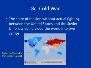
Chapter 8c
Chapter 8c. Neurons: Cellular and Network Properties. Integration: Divergence. Figure 8-25a. Integration: Convergence. Figure 8-25b. Integration: The Abundance of Synapses on a Postsynaptic Neuron. Axon terminals of presynaptic neurons. Dendrite of postsynaptic neuron.


Chapter 8c
E N D
Presentation Transcript
Chapter 8c Neurons: Cellular and Network Properties
Integration: Divergence Figure 8-25a
Integration: Convergence Figure 8-25b
Integration: The Abundance of Synapses on a Postsynaptic Neuron Axon terminalsof presynapticneurons Dendrite ofpostsynapticneuron Glial cellprocesses Axon Figure 8-26
Integration: Purkinje cell • The highly branched dendrites of a Purkinje cell (neuron) demonstrate convergence Figure 8-27
Integration: Spatial Summation Presynaptic axon terminal 1 Three excitatory neurons fire. Their graded potentials separately are all below threshold. 1 T rigger zone Action potential (a) Figure 8-28a, step 1
Integration: Spatial Summation Presynaptic axon terminal 1 Three excitatory neurons fire. Their graded potentials separately are all below threshold. 1 2 Graded potentials arrive at trigger zone together and sum to create a suprathreshold signal. 2 T rigger zone Action potential (a) Figure 8-28a, step 2
Integration: Spatial Summation Presynaptic axon terminal 1 Three excitatory neurons fire. Their graded potentials separately are all below threshold. 1 2 Graded potentials arrive at trigger zone together and sum to create a suprathreshold signal. 3 An action potential is generated. 2 T rigger zone 3 Action potential (a) Figure 8-28a, step 3
Integration: Spatial Summation Presynaptic axon terminal 1 Three excitatory neurons fire. Their graded potentials separately are all below threshold. 1 2 Graded potentials arrive at trigger zone together and sum to create a suprathreshold signal. 3 An action potential is generated. 2 T rigger zone 3 Action potential (a) Figure 8-28a
Integration: Spatial Summation 1 One inhibitory and two excitatory neurons fire. Inhibitory neuron 1 Trigger zone No action potential (b) Figure 8-28b, step 1
Integration: Spatial Summation 1 One inhibitory and two excitatory neurons fire. 2 The summed potentials are below threshold, so no action potential is generated. Inhibitory neuron 1 2 Trigger zone No action potential (b) Figure 8-28b, step 2
Integration: Spatial Summation 1 One inhibitory and two excitatory neurons fire. 2 The summed potentials are below threshold, so no action potential is generated. Inhibitory neuron 1 2 Trigger zone No action potential (b) Figure 8-28b
Integration: Temporal Summation Figure 8-29a
Integration: Temporal Summation Figure 8-29b
3 2 1 3 2 1 Integration: Presynaptic Inhibition (a) Presynaptic inhibition No neurotransmitterrelease Inhibitory neuron Target cell Presynapticaxon terminal No response Excitatoryneuron Response Action potential Neurotransmitterreleased Response An excitatory neuronfires. An action potentialis generated. An inhibitory neuron fires, blockingneurotransmitter release at one synapse. Figure 8-31a
Integration: Postsynaptic Inhibition (b) Postsynaptic inhibition No response Inhibitory neuron modulates the signal. IPSP + No response EPSP Excitatoryneuron No response One excitatory and oneinhibitory presynapticneuron fire. Modulated signal inpostsynaptic neuronbelow threshold. No action potentialinitiated at trigger zone. 1 2 3 No response inany target cell. 4 PLAY Interactive Physiology® Animation: Nervous II: Synaptic Potentials and Cellular Integration Figure 8-31b
Integration: Growth Cones of a Developing Axon • Survival of neurons depend on neurotrophic factors Figure 8-33
Integration: Injury to Neurons Site of injury Proximal stump Distal stump Axon Myelin Figure 8-34
Summary • Organization of the nervous system • CNS – brain and spinal cord • PNS – peripheral nerves and ganglia, sensory receptors • Afferent – sensory • Efferent motor • Somatic • Autonomic • Autonomic • Sympathetic • Parasympathetic
Summary • Cells of the nervous system • Cell body, dendrites, axon, and axon terminal • Interneurons, synapse, postsynaptic cell, presynaptic cell, synaptic cleft, and axonal transport • Glial cells, Schwann cells, satellite cells, microglial, oligodendrocytes, astrocytes, and ependymal cells • Myelin sheaths, nodes of Ranvier, and neural stem cells
Summary • Electrical signals in neurons • GHK equation, graded potentials, local current flow, action potentials, trigger zone, threshold, and all-or-none depolarizations • Activation gate, inactivation gate, absolute refractory period, relative refractory period, and conduction
Summary • Cell-to-cell communication • Electrical synapses, chemical synapses, and synaptic vesicles • Cholinergic neurons, adrenergic neurons, acetylcholine, norepinephrine, glutamate, GABA, serotonin, adenosine, and nitric oxide • Fast synaptic potentials and slow synaptic potentials • Integration of neural information transfer • Divergence, convergence, spatial summation, temporal summation, presynaptic modulation, postsynaptic modulation, and long-term potentiation