Download
1 / 208
2.08k likes | 2.09k Views
Urine Formation by the Kidneys: Glomerular Filtration, Renal Blood Flow, and Their Control Dr. Mard. The kidneys serve multiple functions, including the following: Excretion of metabolic waste products and foreign chemicals Regulation of water and electrolyte balances

E N D
Urine Formation by the Kidneys: Glomerular Filtration, Renal Blood Flow, and Their Control Dr. Mard
The kidneys serve multiple functions, including the following: • Excretion of metabolic waste products and foreign chemicals • Regulation of water and electrolyte balances • Regulation of body fluid osmolality and electrolyte concentrations • Regulation of arterial pressure • Regulation of acid-base balance • Secretion, metabolism, and excretion of hormones • Gluconeogenesis
Excretion of metabolic waste products and foreign chemicals : include • urea(from the metabolism of amino acids) • creatinine(from muscle creatine), • uric acid(from nucleic acids) • end products of hemoglobin breakdown (such as bilirubin) • metabolites of various hormones • most toxins and other foreign substances such as pesticides, drugs, and food additives.
Regulation of Water and Electrolyte Balances. For maintenance of homeostasis, excretion of water and electrolytes must precisely match intake. Intake = Output (excretion) Figure 26-1 Effect of increasing sodium intake 10-fold (from 30 to 300 mEq/day) on urinary sodium excretion and extracellular fluid volume. The shaded areas represent the net sodium retention or the net sodium loss, determined from the difference between sodium intake and sodium excretion.
Regulation of Arterial Pressure Long-term regulation of arterial pressure by excreting variable amounts of sodium and water. Short-term arterial pressure regulation by secreting vasoactive factors or substances, such as Renin angiotensin II
Regulation of Acid-Base Balance The kidneys contribute to acid-base regulation, along with the lungs and body fluid buffers, by excreting acids and by regulating the body fluid buffer stores. The kidneys are the only means of eliminating from the body certain types of acids, such as sulfuric acid and phosphoric acid, generated by the metabolism of proteins.
Regulation of Erythrocyte Production Hypoxia erythropoietin RBC production CRF (hemodialysis) Erythropoietin Anaemia
The kidneys produce the active form of vitamin D, 1,25-dihydroxyvitamin D3 (calcitriol), by hydroxylating this vitamin at the "number 1" position.
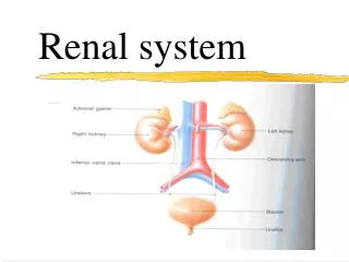
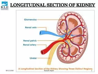
Physiologic Anatomy of the Kidneys Location : posterior wall of the abdomen (retroperitoneal) Weight : 150 gr Hilum : entrance of renal pedicles and ureter Fibrous capsule :The kidney is surrounded by a tough, that protects its delicate inner structures.
The kidney composed of : two major regions that can be visualized are the outer cortexand the inner region (medulla). Renal pyramids : The medulla is divided into multiple cone-shaped masses of tissue called renal pyramids. The base of each pyramid originates at the border between the cortex and medulla and terminates in the papilla, which projects into the space of the renal pelvis. Renal pelvis: a funnel-shaped continuation of the upper end of the ureter. The outer border of the pelvis is divided into open-ended pouches called major calyces that extend downward and divide into minor calyces
Minor calyces : collect urine from the tubules of each papilla. contractile elements : The walls of the calyces, pelvis, and ureter contain contractile elements that propel the urine toward the bladder
Renal Blood Supply Blood flow: is normally about 22% of the C.O or 1100 ml/min. The renal artery enters the kidney through the hilum and then branches progressively to form : Afferent arterioles glomerular capillaries Efferent arteriole peritubular capillaries
The renal circulation is unique in that it has two capillary beds, the glomerular and peritubular capillaries, which are arranged (in series), which help regulate the hydrostatic pressure in both sets of capillaries. • High hydro-static pressure in the glomerular capillaries (60 mm Hg) causes rapid fluid filtration, • Whereas a much lower hydrostatic pressure in the peritubular capillaries (about 13 mm Hg) permits rapid fluid reabsorption. • By adjusting the resistance of the afferent and efferent arterioles, the kidneys can regulate the hydrostatic pressure in both the glomerular and the peritubular capillaries, thereby changing the rate of glomerular filtration, tubular reabsorption
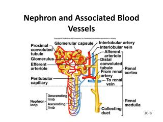
The Nephron Is the Functional Unit of the Kidney Each kidney (1 million nephrons) Each nephrons capable of forming urine. The kidney cannot regenerate new nephrons Therefore, with renal injury, disease, or normal aging, there is a gradual decrease in nephron number. After age 40, the number of functioning nephrons usually decreases about 10 per cent every 10 years. Each nephron contains : • The glomerulus • a long tubule in which the filtered fluid is converted into urine on its way to the pelvis of the kidney
The glomerulus contains a network of branching and anastomosing glomerular capillaries that, compared with other capillaries, have high hydrostatic pressure (60 mm Hg). The glomerular capillaries are covered by epithelial cells, and the total glomerulus is encased in Bowman's capsule. Fluid filtered from the glomerular capillaries flows into Bowman's capsule and then into the proximal tubule
Pain sensation in ureter • Reflex. The ureters are well supplied with pain nerve • fibers. When a ureter becomes blocked (e.g., by a ureteral stone), intense reflex constriction occurs, which is associated with severe pain. Also, the pain impulses cause a sympathetic reflex back to the kidney to constrict the renal arterioles, thereby decreasing urine output from the kidney. This effect is called the ureterorenal reflex and is important for preventing excessive flow of fluid into the pelvis of a kidney with a blocked ureter.
Innervation of the Bladder. • The principal nerve supply of the bladder-the pelvic nerves, which connect with the spinal cord through the sacral plexus, (S2 and S3) • The sensory fibers detect the degree of stretch in the bladder wall. • Stretch signals from the posterior urethra are especially strong and are mainly responsible for initiating the reflexes that cause bladder emptying. • The motor nerves transmitted in the pelvic nerves are parasympathetic fibers. • In addition to the pelvic nerves, two other types of innervation are important in bladder function. Most important are the skeletal motor fibers transmitted through the pudendal nerve to the external bladder sphincter. • These fibers are somatic nerve fibers that innervate and control the voluntary skeletal muscle of the sphincter. • Also, the bladder receives sympathetic innervation from the sympathetic chain through the hypogastric nerves, connecting mainly with the L2 segment of the spinal cord. These sympathetic fibers stimulate mainly the blood vessels and have little to do with bladder contraction. • Some sensory nerve fibers also pass by way of the sympathetic nerves and may be important in the sensation of fullness and, in some instances, pain.
Figure 26-8 shows the approximate changes in intravesicular pressure as the bladder fills with urine. When there is no urine in the bladder, the intravesicular pressure is about 0, but by the time 30 to 50 milliliters of urine have collected, the pressure rises to 5 to 10 centimeters of water. • Additional urine—200 to 300 milliliters—can collect with only a small additional rise in pressure; this constant level of pressure is caused by intrinsic tone of the bladder wall. Beyond 300 to 400 milliliters, collection of more urine in the bladder causes the pressure to rise rapidly. • Superimposed on the tonic pressure changes during filling of the bladder are periodic acute increases in pressure that last from a few seconds to more than a minute. The pressure peaks may rise only a few centimeters of water or may rise to more than 100 centimeters of water. These pressure peaks are called micturition waves in the cystometrogram and are caused by the micturition reflex.
MICTURITION REFLEX • As the bladder fills, many superimposed micturition contractions begin to appear, as shown by the dashed spikes. They are the result of a stretch reflex initiated by sensory stretch receptors in the bladder wall, especially by the receptors in the posterior urethra when this area begins to fill with urine at the higher bladder pressures. • Sensory signals from the bladder stretch receptors are conducted to the sacral segments of the cord through the pelvic nerves and then reflexively back again to the bladder through the parasympathetic nerve fibers by way of these same nerves. • When the bladder is only partially filled, these micturition contractions usually relax spontaneously after a fraction • of a minute, the detrusor muscles stop contracting, and pressure falls back to the baseline. • As the bladder continues to fill, the micturition reflexes become more frequent and cause greater contractions of the detrusor muscle. • Once a micturition reflex begins, it is “self-regenerative.” That is, initial contraction of the bladder activates the • stretch receptors to cause a greater increase in sensory impulses from the bladder and posterior urethra, which • causes a further increase in reflex contraction of the bladder; thus, the cycle is repeated again and again until • the bladder has reached a strong degree of contraction. • Once the micturition reflex becomes powerful enough, it causes another reflex, which passes through the pudendal • nerves to the external sphincter to inhibit it. If this inhibition is more potent in the brain than the voluntary • constrictor signals to the external sphincter, urination will occur. If not, urination will not occur until the bladder fills still further and the micturition reflex becomes more powerful.
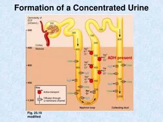
Macula densa: At the end of the thick ascending limb is a short segment, which is actually a plaque in its wall, known as the macula densa • Distal tubule : Beyond the macula densa, fluid enters the distal tubule, which, like the proximal tubule, lies in the renal cortex. This is followed by the connecting tubule and the cortical collecting tubule, which lead to the cortical collecting duct. • The initial parts of 8 to 10 cortical collecting ducts join to form a single larger collecting duct that runs downward into the medulla and becomes the medullary collecting duct. • The collecting ducts merge to form progressively larger ducts that eventually empty into the renal pelvis through the tips of the renal papillae. • In each kidney, there are about 250 of the very large collecting ducts, each of which collects urine from about 4000 nephrons.
Regional Differences in Nephron Structure (Length of Henle & the vascular structures) Cortical : 1. Glomeruli located in the outer cortex 2. They have short loops of Henle that penetrate only a short distance into the medulla 3. The entire tubular system is surrounded by an extensive network of peritubular capillaries Juxtamedullary Nephrons (20 to 30 % of the nephrons) : 1. glomeruli that lie deep in the renal cortex near the medulla 2.These nephrons have long loops of Henle that dip deeply into the medulla 3. long efferent arterioles extend from the glomeruli down into the outer medulla and then divide into specialized peritubular capillaries called vasa recta (this specialized network of capillaries in the medulla plays an essential role in the formation of a concentrated urine).
Urine Formation Results from Glomerular Filtration, Tubular Reabsorption, and Tubular Secretion • The rates at which different substances are excreted in the urine represent the sum of three renal processes, shown in • (1) glomerular filtration • (2) reabsorption • (3) secretion Expressed mathematically: Urinary excretion rate= Filtration rate- Reabsorption rate + Secretion rate
Urinary excretion rate= Filtration rate- Reabsorption rate + Secretion rate
Ultrafiltation : • Urine formation begins when a large amount of fluid that is virtually free of protein is filtered from the glomerular capillaries into Bowman's capsule. Most substances in the plasma, except for proteins, are freely filtered, so that their concentration in the glomerular filtrate in Bowman's capsule is almost the same as in the plasma.
Panel Ais freely filtered by the glomerular capillaries but is neither reabsorbed nor secreted. Therefore, its excretion rate is equal to the rate at which it was filtered (such as creatinine) Panel B, the substance is freely filtered but is also partly reabsorbed from the tubules back into the blood (rate of urinary excretion < rate of filtration): electrolytes In this case, the excretion rate is calculated as the filtration rate minus the reabsorption rate. Panel C, the substance is freely filtered at the glomerular capillaries but is not excreted (comp[letly absorbed) shuch as : nutritional substances (amino acids and glucose) Panel D is freely filtered at the glomerular capillaries and is not reabsorbed, but additional quantities of this substance are secreted from the peritubular capillary blood into the renal tubules (organic acids and bases. The excretion rate in this case is calculated as filtration rate plus tubular secretion rate.
Filtration, Reabsorption, and Secretion of Different Substances In general, tubular reabsorption is quantitatively more important than tubular secretion in the formation of urine, but secretion plays an important role in determining the amounts of K and H ions Most substances that must be cleared from the blood, especially the end products of metabolism such as urea, creatinine, uric acid, and urates, are poorly reabsorbed and are therefore excreted in large amounts in the urine.
Why Are Large Amounts of Solutes Filtered and Then Reabsorbed by the Kidneys? One might question the wisdom of filtering such large amounts of water and solutes and then reabsorbing most of these substances. • One advantage of a high GFR is that it allows the kidneys to rapidly remove waste products from the body that depend primarily on glomerular filtration for their excretion. 2. A second advantage of a high GFR is that it allows all the body fluids to be filtered and processed by the kidney many times each day. Because the entire plasma volume is only about 3 liters, whereas the GFR is about 180 L/day, the entire plasma can be filtered and processed about 60 times each day. This high GFR allows the kidneys to precisely and rapidly control the volume and composition of the body fluids.
Glomerular Filtration-The First Step in Urine Formation Filtrate composition s Free from proteins and devoid of cellular elements, including red blood cells. The concentrations of other constituents of the glomerular filtrate, including most salts and organic molecules, are similar to the concentrations in the plasma. Exceptions for calcium and fatty acids because of almost one half of the plasma calcium and most of the plasma fatty acids are bound to proteins
GFR Is About 20 % of the Renal Plasma Flow GFR is determined by: • P and π • the capillary filtration coefficient (Kf) [the product of the permeability x A] Kf of glomerular capillaries=400 other capillaries
In the average adult human, the GFR is about 125 ml/min, or 180 L/day. The fraction of the renal plasma flow that is filtered (the filtration fraction) averages about 0.2; this means that about 20 % of the plasma flowing through the kidney is filtered through the glomerular capillaries Filtration fraction : GFR/Renal plasma flow
Glomerular Capillary Membrane The glomerular capillary membrane is similar to that of other capillaries, except that it has three (instead of the usual two) major layers: • Endothelium • A basement membrane • A layer of epithelial cells (podocytes) Together, these layers make up the filtration barrier, which, despite the three layers, filters several hundred times as much water and solutes as the usual capillary membrane. Ultra-filtration : Even with this high rate of filtration, the glomerular capillary membrane normally prevents filtration of plasma proteins.
Filterability of Solutes Is Inversely Related to Their Size The glomerular capillary membrane is thicker than most other capillaries, but it is also much more porous and therefore filters fluid at a high rate. Despite the high filtration rate, the glomerular filtration barrier is selective in determining which molecules will filter, based on their size and electrical charge.
Negatively Charged Large Molecules Are Filtered Less Easily Than Positively Charged Molecules of Equal Molecular Size • The molecular diameter of the plasma protein albumin is only about 6 nm, whereas the pores of the glomerular membrane are thought to be about 8 nanometers • Albumin is restricted from filtration, however, because of its negative charge and the electrostatic repulsion exerted by negative charges of the glomerular capillary wall proteoglycans
In certain kidney diseases, the negative charges on the basement membrane are lost even before there are noticeable changes in kidney histology, a condition referred to as minimal change nephropathy. As a result of this loss of negative charges on the basement membranes, some of the lower-molecular-weight proteins, especially albumin, are filtered and appear in the urine, a condition known as proteinuria or albuminuria.
Determinants of the GFR GFR is determined by • the sum of the hydrostatic and colloid osmotic forces across the glomerular membrane, which gives the net filtration pressure, and • the glomerular capillary filtration coefficient, Kf. Expressed mathematically, the GFR equals the product of Kf and the net filtration pressure: GFR = Kf * Net filtration pressure
The net filtration pressure represents the sum of the hydrostatic and colloid osmotic forces that either favor or oppose filtration across the glomerular capillaries . • These forces include (1) hydrostatic pressure inside the glomerular capillaries (glomerular hydrostatic pressure, PG), which promotes filtration • (2) the hydrostatic pressure in Bowman's capsule (PB) outside the capillaries, which opposes filtration • (3) the colloid osmotic pressure of the glomerular capillary plasma proteins (πG), which opposes filtration • (4) the colloid osmotic pressure of the proteins in Bowman's capsule (πB), which promotes filtration. (Under normal conditions, the concentration of protein in the glomerular filtrate is so low that the colloid osmotic pressure of the Bowman's capsule fluid is considered to be zero.) • The GFR can therefore be expressed as
Increased Glomerular Capillary Filtration Coefficient Increases GFR
Increased Bowman's Capsule Hydrostatic Pressure Decreases GFR In certain pathological states associated with obstruction of the urinary tract, Bowman's capsule pressure can increase markedly, causing serious reduction of GFR. For example, precipitation of calcium or of uric acid may lead to "stones" that lodge in the urinary tract, often in the ureter, thereby obstructing outflow of the urinary tract and raising Bowman's capsule pressure. This reduces GFR and eventually can damage or even destroy the kidney unless the obstruction is relieved.