A 'normal' resting coronary heart rate will not be so normal in spite of everything
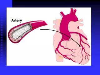
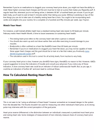
Tachycardia is mostly thought-about to be a resting heart fee of over a hundred bpm, based on theNational Institutes of Health, and usually triggered when electrical indicators within the coronary heart's higher chambers fire abnormally. If the center price is nearer to one hundred fifty bpm or greater, it's a situation often known as supraventricular tachycardia (SVT). <h3>Watch Your Heart</h3> To velocity up, one need merely remove one's foot from the brake and let the engine improve velocity. In the case of the center, reducing parasympathetic stimulation decreases the release of ACh, which allows HR to increase up to roughly 100 bpm. Any increases beyond this price would require sympathetic stimulation. The heart rate is rhythmically generated by the sinoatrial node. It can also be influenced by central factors by way of sympathetic and parasympathetic nerves. <h3>RELATED:u00c2u00a0Everyday Ways to Help Your Heart</h3> <ul><li>You also needs to warm up and cool down earlier than and after exercising to avoid injury to your muscle tissue.</li><li>Bradycardia is typically outlined as a coronary heart rate thatu00e2u20acu2122s lower than 60 beats per minute.</li><li>Remember if you're on drugs to slow your coronary heart price down, you may not be capable of meet these upper coronary heart rates and the goal should be to exercise at a fee that makes you lightly puff.</li><li>The calculation used in this device is correct for all exercise levels, from inactive to very active.</li></ul> Nervous affect over the heart rate is centralized throughout the two paired cardiovascular centres of the medulla oblongata. During rest, both facilities provide slight stimulation to the guts, contributing to autonomic tone. Normally, vagal stimulation predominates as, left unregulated, the SA node would provoke a sinus rhythm of approximately 100 bpm. This tool will help you discover your target heart fee based mostly in your age, resting heart rate, and activity degree. <h2>RELATED:u00c2u00a0Five Tips for A Healthy Heart</h2> Without any nervous stimulation, the SA node would set up a sinus rhythm of approximately one hundred bpm. Since resting rates are considerably less than this, it turns into evident that parasympathetic stimulation normally slows HR. This is just like a person driving a car with one foot on the brake pedal. Your resting coronary heart fee refers back to the number of beats your coronary heart pumps if youu00e2u20acu2122re not exerting yourselfu00e2u20acu201dlike when youu00e2u20acu2122re relaxed, lying down, and calm. Your coronary heart price is often lowest when sleeping or in any other case inactive and then increases with bodily exercise. Your coronary heart fee dictates the precise number of occasions your coronary heart beats per minute.
26 views • 1 slides