Download

1 / 52
520 likes | 558 Views
Electrophoresis is a method where charged molecules migrate in response to an electrical field, used analytically to study properties of molecules or as a separating technique. Gel units such as tube, slab, or flat bed are commonly used, with electrical parameters like resistance, voltage, current, and power playing key roles. The net charge of molecules is influenced by the pH of the medium, and temperature control is crucial during electrophoresis stages. The solid support matrix aids in separation and results staining. Agarose and polyacrylamide gels are utilized for their unique porous properties, allowing for effective molecular sieving based on size differences. Agarose gels are fragile, while polyacrylamide gels are tougher due to covalent crosslinking. Key considerations for polyacrylamide gel electrophoresis include pore size determination, acrylamide percentage adjustment, crosslinker variation, and proper gel preparation techniques.

E N D
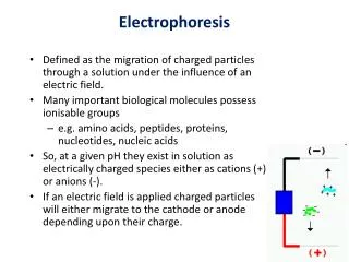
Electrophoresis • Electrophoresis is a method whereby charged molecules in solution, chiefly proteins and nucleic acids, migrate in response to an electrical field. • Their rate of migration through the electrical field, depends on the strength of the field, on the net charge, size, and shape of the molecules, and also on the ionic strength, viscosity, and temperature of the medium in which the molecules are moving. • As an analytical tool, electrophoresis is simple, rapid and highly sensitive. • It can be used analytically to study the properties of a single charged species or mixtures of molecules. It can also be used preparatively as a separating technique
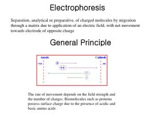
Electrophoresis • Electrophoresis is usually done with gels formed in tubes, slabs, or on a flat bed. • In many electrophoresis units, the gel is mounted between two buffer chambers containing separate electrodes, so that the only electrical connection between the two chambers is through the gel.
In most electrophoresis units, the gel is mounted between two buffer chambers containing separate electrodes so that the only electrical connection between the two chambers is through the gel.
Interrelation of Resistance, Voltage, Current and Power • Two basic electrical equations are important in electrophoresis • The first is Ohm's Law, I = E/R • The second is P = EI • This can also be expressed as P = I2R • In electrophoresis, one electrical parameter, either current, voltage, or power, is always held constant
Consequences • Under constant current conditions (velocity is directly proportional to current), the velocity of the molecules is maintained, but heat is generated. • Under constant voltage conditions, the velocity slows, but no additional heat is generated during the course of the run • Under constant power conditions, the velocity slows but heating is kept constant
The Net Charge is Determined by the pH of the Medium • Proteins are amphoteric compounds, that is, they contain both acidic and basic residues • Each protein has its own characteristic charge properties depending on the number and kinds of amino acids carrying amino or carboxyl groups • Nucleic acids, unlike proteins, are not amphoteric. They remain negative at any pH used for electrophoresis
Temperature and Electrophoresis • Important at every stage of electrophoresis • During Polymerization • Exothermic Reaction • Gel irregularities • Pore size • During Electrophoresis • Denaturation of proteins • Smile effect • Temperature Regulation of Buffers
What is the Role of the Solid Support Matrix? • It inhibits convection and diffusion, which would otherwise impede separation of molecules • It allows a permanent record of results through staining after run • It can provide additional separation through molecular sieving
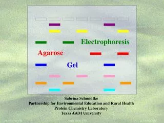
Agarose and Polyacrylamide • Although agarose and polyacrylamide differ greatly in their physical and chemical structures, they both make porous gels. • A porous gel acts as a sieve by retarding or, in some cases, by completely obstructing the movement of macromolecules while allowing smaller molecules to migrate freely. • By preparing a gel with a restrictive pore size, the operator can take advantage of molecular size differences among proteins
Agarose and Polyacrylamide • Because the pores of an agarose gel are large, agarose is used to separate macromolecules such as nucleic acids, large proteins and protein complexes • Polyacrylamide, which makes a small pore gel, is used to separate most proteins and small oligonucleotides. • Both are relatively electrically neutral
Agarose Gels • Agarose is a highly purified uncharged polysaccharide derived from agar • Agarose dissolves when added to boiling liquid. It remains in a liquid state until the temperature is lowered to about 40° C at which point it gels • The pore size may be predetermined by adjusting the concentration of agarose in the gel • Agarose gels are fragile, however. They are actually hydrocolloids, and they are held together by the formation of weak hydrogen and hydrophobic bonds
Structure of the Repeating Unit of Agarose, 3,6-anhydro-L-galactose Basic disaccharide repeating units of agarose, G: 1,3-β-d-galactose and A: 1,4-α-l-3,6-anhydrogalactose
Polyacrylamide Gels • Polyacrylamide gels are tougher than agarose gels • Acrylamide monomers polymerize into long chains that are covalently linked by a crosslinker • Polyacrylamide is chemically complex, as is the production and use of the gel
Considerations with PAGE • Preparing and Pouring Gels • Determine pore size • Adjust total percentage of acrylamide • Vary amount of crosslinker • Remove oxygen from mixture • Initiate polymerization • Chemical method • Photochemical method
Considerations with PAGE • Analysis of Gel • Staining or autoradiography followed by densitometry • Blotting to a membrane, either by capillarity or by electrophoresis, for nucleic acid hybridization, autoradiography or immunodetection
SDS Gel Electrophoresis • In SDS separations, migration is determined not by intrinsic electrical charge of polypeptides but by molecular weight • Sodium dodecylsulfate (SDS) is an anionic detergent which denatures secondary and non–disulfide–linked tertiary structures by wrapping around the polypeptide backbone. In so doing, SDS confers a net negative charge to the polypeptide in proportion to its length • When treated with SDS and a reducing agent, the polypeptides become rods of negative charges with equal “charge densities" or charge per unit length.
Continuous and Discontinuous Buffer Systems • A continuous system has only a single separating gel and uses the same buffer in thetanks and the gel • In a discontinuous system a nonrestrictive large pore gel, called a stacking gel, is layered on top of a separating gel • The resolution obtainable in a discontinuous system is much greater than that obtainable in a continuous one. However, the continuous system is a little easier to set up
Determining Molecular Weights of Proteins by SDS-PAGE • Run a gel with standard proteins of known molecular weights along with the polypeptide to be characterized • A linear relationship exists between the log10 of the molecular weight of a polypeptide and its Rf • Rf = ratio of the distance migrated by the molecule to that migrated by a marker dye-front • The Rf of the polypeptide to be characterized is determined in the same way, and the log10 of its molecular weight is read directly from the standard curve
Blotting • Blotting is used to transfer proteins or nucleic acids from a slab gel to a membrane such as nitrocellulose, nylon, DEAE, or CM paper • The transfer of the sample can be done by capillary or Southern blotting for nucleic acids (Southern, 1975) or by electrophoresis for proteins or nucleic acids
Isoelectric Point • There is a pH at which there is no net charge on a protein; this is the isoelectric point (pI). • Above its isoelectric point, a protein has a net negative charge and migrates toward the anode in an electrical field. • Below its isoelectric point, the protein is positive and migrates toward the cathode.
Isoelectric Focusing • Isoelectric focusing is a method in which proteins are separated in a pH gradient according to their isoelectric points • Focusing occurs in two stages; first, the pH gradient is formed • In the second stage, the proteins begin their migrations toward the anode if their net charge is negative, or toward the cathode if their net charge is positive • When a protein reaches its isoelectric point (pI) in the pH gradient, it carries a net charge of zero and will stop migrating
Two-Dimensional Gel Electrophoresis • Two-dimensional gel electrophoresis is widely used to separate complex mixtures of proteins into many more components than is possible in conventional one-dimensional electrophoresis • Each dimension separates proteins according to different properties
O’Farrell 2D Gel System • The first dimension tube gel is electrofocused • The second dimension is an SDS slab gel • The analysis of 2-D gels is more complex than that of one-dimensional gels because the components that show up as spots rather than as bands must be assigned x, y coordinates
DIGE DIfference Gel Electrophoresis • DIGE can be done in one-or two-dimensions. Same principle. • Requires fluorescent protein stains (up to three of these), a gel box, and a gel scanner. • Dyes include Cy2, Cy3 and Cy5 (Amersham system). • These have similar sizes and charges, which means that individual proteins move to the same places on 2-D gels no matter what dye they are labeled with. • Detection down to 125 pg of a single protein.
Linear response to protein concentration over a 105 concentration range. • Standard chemistry links dyes to proteins via lysine side-chains. • Proteins samples are produced by homogenizing cells.
After running the gels, three scans are done to extract the Cy2, Cy3, and Cy5 fluorescence values. • Assuming the Cy2 is the internal control, this is used to identify and positionally match all spots on the different gels. • The intensities are then compared for the Cy3 and Cy5 values of the different spots, and statistics done to see which ones have significantly changed in intensity as a consequence of the experimental treatment.
Labeling slightly shifts the masses of the proteins, so to cut them out for further analysis, you first stain the gel with a total protein stain. • SYPRO Ruby is used for this purpose (Molecular Probes). • When designing 2-D DIGE experiments, the following recommendations should be considered: • Inclusion of an internal standard sample on each gel. These can comprise a mixture of known proteins of different sizes, or simply a mixture of unknown proteins (one of your samples). • Use of biological replicates. • Randomization of samples to produce unbiased results.