Download

1 / 41
410 likes | 483 Views
Explore the organization of the central and peripheral nervous systems, types of neurons, supportive cells, gray and white matter, and neural tube development.

E N D
Basic Organization of the Nervous System Dr. G.R. Leichnetz
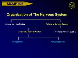
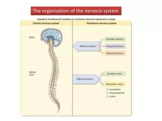
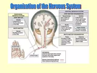
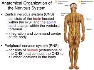
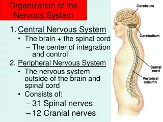
The central nervous system (CNS) consists of the: brain and spinal cord The peripheral nervous system (PNS) consists of: 12 pairs of cranial nerves, 31 pairs of spinal nerves, nerve plexuses, ganglia, and emergent peripheral nerves
Neural tissue of the CNS and PNS contains two general categories of cells: neurons and supportive cells. The central nervous system (CNS) consists of about 10% neurons and 90% neuroglia (supportive cells). The CNS contains approximately 10 billion neurons. While the PNS contains all three morphological types of neurons (unipolar, bipolar, multipolar), the CNS only contains multipolar neurons. These multipolar neurons, however, are of a wide variety of size and shape. The neuroglia (supportive cells) of the CNS are astrocytes, oligodendrocytes, and microglia. The supportive cells of the PNS are Schwann cells and satellite cells.
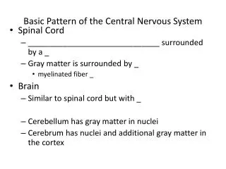
Typical Neuron Gray matter contains cell bodies of neurons. CNS gray matter is organized in two ways: nucleus (cluster) or cortex (layers). Note: A ganglion is a cluster of neurons in the PNS. White matter contains myelinated axons. Both gray matter and white matter also contain neuroglia (astrocytes, oligodendrocytes). Cell Body Myelinated axon From: Noback & Demarest, The Human Nervous System
Cytoarchitecture of the Brain Nuclei contain the cell bodies of neurons Nissl-stained (cresyl-violet) cross section of medulla (see “nuclei,” containing cell bodies, stained darker purple)
Myeloarchitecture of the Brain Tracts contain myelinated axons of neurons Compare to C.V.-stained section Myelin-stained section of medulla (see tracts, containing myelinated axons, stained darker black; nuclei are pale)
A “nucleus” is a cluster of cell bodies of neurons in the CNS. Myelin-stained section through the midbrain, showing the trochlear nucleus(pale round structure just above small dark tract) The neuron cell bodies which make up the trochlear nucleus can be seen by injecting HRP into its target muscle (superior oblique muscle of the eye). The enzyme is retrogradely transported back to parent cell body. The trochlear nucleus seen here contains the cell bodies of many multipolar motor neurons. Trochlear nucleus Darkfield photomicrograph showing HRP-containing cell bodies of neurons
A “cortex” is laminated gray matter. The surface of the cerebrum is covered with gray matter, the cerebral cortex, which contains layers of neuronal cell bodies. The cerebrum and cerebellum each have a cortex. In a coronal section of the brain, the laminated gray matter (cerebral cortex) covers the surface of the cerebrum (Note: subcortical nuclei- black arrows). Cortex Nuclei
Cresyl-violet stained section of the cerebral cortex. In a “cortex” the neurons are in layers on the surface of the cerebrum. The cerebral cortex has six cellular layers. CORTEX Layer III Pyramidal neurons Subcortical white matter
Brain Development: Basic Neuroembryology
The entire CNS develops from the embryonic neural tube. Neural ectoderm on the dorsal aspect of the developing embyro gives rise to neural groove, neural folds, and then fuses to form the neural tube. The neural crest pinches off dorsolateral to the neural tube, and gives rise to neural structures in the PNS.
Neural Tube: gives rise to CNS Neural Crest: gives rise to PNS Both are “neural ectoderm” and contain primordial cells which give rise to neurons and supportive elements (ie. neuroglia in CNS; Schwann cells & satellite cells in PNS)
Rhombencephalon Mesencephalon Prosencephalon Mesencephalon Diencephalon Metencephalon Myelencephalon Telencephalon The rostral end of the neural tube undergoes cephalization (developing brain) giving rise to three primary brain vesicles (prosen-, mesen- and rhomben- cephalon); then five secondary vesicles (telen-, dien-, mesen-, meten-, myelen- cephalon).
The three primary brain vesicles (prosen-, mesen-, and rhomben-cephalon) differentiate into five secondary brain vesicles (telen-, dien-, mesen-, meten-, myelen-cephalon).
The terms for the five embryonic secondary brain vesicles are used in the adult for the five subdivisions of the brain. Five subdivisions of adult brain: Telencephalon Diencephalon Mesencephalon Metencephalon Myelencephalon (Metencephalon) (Mesencephalon) (Myelencephalon)
Brain Development: Differentiation of the Neural Tube
The mantle layer of the neural tube (presumptine gray matter) differentiates into neuroblasts (neurons) and glioblasts (neuroglia). The marginal layer (presumptive white matter) contains processes of developing neurons. The ependymal layer, which is a germinal layer in the embryonic neural tube, lines the neurocoel (ventricles). Mantle layer Ependymal layer Neuroblasts that give rise to neurons do not undergo mitosis PNS The neural tube has three layers. Marginal layer CNS Glioblasts give rise to neuroglia which retain the capacity to divide throughout life From: Noback & Demarest, The Human Nervous System
Differentiation of Neural Tube Into Spinal Cord The mantle layer of the neural tube (presumptive gray matter) gives rise to alar and basal plates, separated by the sulcus limitans. The alar plate differentiates into sensory nuclei of the CNS (dorsal horn of the spinal cord gray); the basal plate into motor nuclei (ventral horn of spinal gray).
Differentiation of Neural Tube Into Brainstem (Medulla) At the level of the developing medulla, the alar plate of the neural tube (which gives rise to sensory nuclei) is displaced laterally, separated from the basal plate (motor nuclei) by the sulcus limitans.
Further differentiation of the alar and basal plates, dividing into cell columns that ultimately break up into sensory and motor nuclei. Cell columns in developing medulla
The longitudinal cell columns (derived from alar and basal plates) break up into sensory and motor nuclei, corresponding to functional components of cranial nerves (sensory: GSA, GVA, SSA, SVA; motor: GSE, GVE, SVE).
Dorsal view of the adult brainstem, showing location of sensory (blue) and motor (red) nuclei, which are derived from sensory and motor cell columns. The longitudinal cell columns break up into separate nuclei. Motor nuclei derived from cell columns that originated from basal plate Sensory nuclei derived from cell columns that originated from alar plate Dorsal view of brainstem with cerebellum removed The sulcus limitans remains as a landmark in the floor of the fourth ventricle, separating motor (medial) and sensory (lateral) nuclei.
Basic LM Neurohistology: Neurons
Morphological Classification of Neurons Unipolar- General sensory- found in cranial and dorsal root ganglia (PNS) Bipolar- Special sensory- found in cochlear & vestibular ganglia, olfactory epithelium, and retina (PNS) Multipolar- All CNS neurons are multipolar. Multipolar neurons in the PNS are found in autonomic ganglia
Dorsal root ganglion containing (pseudo-) unipolar neurons Dorsal root ganglia and cranial sensory ganglia contain unipolar neuron cell bodies. Origin of single process Unipolar neurons in the dorsal root ganglion (derived from neural crest) are associated with general sensation (eg. pain, temp., touch, and visceral sense). The small nuclei between neuronal cell bodies are satellite cells and fibroblasts. Unipolar neuron cell body
Bipolar neurons are found in ganglia associated with special senses (eg. cochlear & vestibular ganglia). The retina also contains bipolar neurons. Bipolar neuron cell body Central process Peripheral process
Multipolar motor neurons of the ventral horn of the spinal cord stained with a silver stain. Neuron cell body Origin of axon (axon hillock) Neuron cell body Neuron cell body Dendrites Origin of axon (axon hillock) Motor neurons of the ventral horn of the spinal cord stained with cresyl violet or H & E
Pyramidal Neurons of the Cerebral Cortex- are multipolar Cresyl Violet (Nissl) Stain Oil 100 X Apical dendrite Silver Stain Basal dendrites H & E Stain Axon Oil 100 X
Purkinje cells of the cerebellar cortex are multipolar neurons. Dendritic arborization H & E Primary dendrite Silver Stain
Basic LM Neurohistology: Neuroglia
Neuroglia of the Central Nervous System: Astrocytes, Oligodendrocytes, Microglia Astrocyte Neuron Astrocyte Haines
Neuroglia support the function of neurons in the CNS Astrocyte Astrocytes: 1. Carry nutrients from capillaries to neurons 2. Maintain optimal ionic conditions around neurons (remove excess neurotransmitter, ions) 3. Neural development- axon guidance, enhance synapse formation 4. Enhance synaptic transmission (uptake calcium, release ATP) Oligodendrocytes: myelination of CNS axons Oligodendrocyte Astrocyte From: Noback & Demarest, The Human Nervous System
Protoplasmic astrocytes are found in gray matter. Protoplasmic astrocyte Pyramidal neuron
Astrocytes have long processes that extend to brain capillaries, ending in perivascular end feet. Capillary Astrocyte Astrocyte Capillary Astrocyte Capillary
Blood-Brain Barrier Selective permeability of the capillary endothelium, excludes certain substances. In the strictest sense, the blood-brain barrier is formed by the tight junctions between capillary endothelial cells. However, some people refer to the blood-brain barrier more inclusively as including the endothelium, basal lamina, and astrocytic end feet.
Myelination of CNS and PNS Axons CNS axons are myelinated by oligodendocytes PNS axons are myelinated by Schwann cells Oligodendrocytes CNS PNS Schwann cells Oligodendrocytes can myelinate as many as 50 internodal segments, whereas Schwann cell myelinate only 1 internode.