The Hypothalamus
350 likes | 812 Views
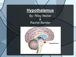
The Hypothalamus. A crucial part of the CNS that takes some part in regulating most organs. 3 major functions (we will review 2 today). Regulating release of hormones from pituitary gland. Regulating the ANS; i.e ., general visceral motor functions we reviewed last time.

The Hypothalamus
E N D
Presentation Transcript
A crucial part of the CNS that takes some part in regulating most organs • 3 major functions (we will review 2 today). • Regulating release of hormones from pituitary gland. • Regulating the ANS; i.e., general visceral motor functions we reviewed last time. • Regulating the “appetitive behaviours” (eating, drinking, mating).
The 3 functional zones of the hypothalamus and the nuclei contained therein. • Regulation of Pituitary: Parvocellular (anterior) ad magnocellular (posterior) neurosecretory systems. • Overview of ANS functional anatomy (sympathetic, parasympathetic systems). • Regulation of autonomic functions by descending projections from the hypothalamus. • Regional anatomy. A. Anterior-posterior sections of hypothalamus and review key nuclei. B. Descending pathway and sc nuclei. C. Clinical Note: Horner’s Syndrome.
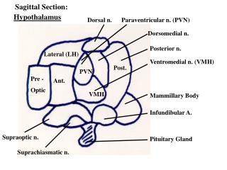
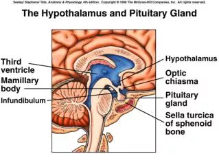
I. 3 Functional Zones • General location of hypothalamus: - ventral to thalamus - just over optic chiasm and pituitary stalk (infundibulum). - divided in half by 3rd ventricle
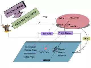
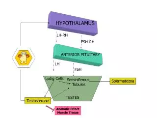
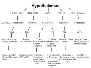
Periventricular zone - a thin nuclei bordering the 3rd ventricle. - regulates release of endocrine hormones from anterior pituitary gland (See Table 15-1). -uses neurosecretion as a portal vein system, rather than a neurotransmitter across a synapse. • Middle zone - regulates hormone release from posterior pituitary. - regulates ANS. • Lateral zone - integration and transmission of info from limbic system structures (important in emotional regulation – will view next lecture (limbic system).
3 Functional hypothalamic zones(Fig. 15-14) – Mediolateral zones
II. Regulation of Pituitary: Parvocellular and Magnocellular Neurosecretory Systems • Parvocellular system and the anterior pit. - Small-diameter neurons in several hypothalamic nuclei (of periventricular zone) – most medial – regulate anterior pituitary hormone release by neurovascular rather than synaptic transmission.
Parvocellular System (Fig. 15-4A) Note the various nuclei
Neurosecretion and Portal Vein System(Fig. 15-5): Note the path: Parvocellular neurosecretory cells anterior lobe via portal vein. Chemicals released are peptides, which either promote or inhibit the release of hormones from anterior lobe secretory cells (Table 15-1).
Magnocellular system and the posterior pituitary. - Here, peptide hormones are produced by large-diameter hypothalamic neurons from same nuclei of the middle zone. - Axons deliver these hormones down the infundibular stalk and terminate on fenestral capillaries (“leaky”) of the posterior pit - this is 1 place lacking a BBB.
Magnocellular System (Fig. 15-4B) Note the paraventricular and supraoptic nuclei Hormones: Vasopressin (ADH) – peptide which incr bp by its effects on vascular smooth muscle as well as by promoting H2O reabsorption from DCTs of kidneys to decr urine vol. Oxytocin – incr uterine contraction and milk ejection from mammary glands.
III. Overview of Autonomic Nervous System Sympathetic and Parasympathetic systems – Fig. 15-7. Clearly distinct anatomical locations of preganglionic (central) neurons. Sympathetic: T1 L3 Parasympathetic: brainstem nuclei (reviewed last time): S2 S4 (sacral spinal cord). Also different locations of post-ganglionic neurons.
Sympathetic: peripheral ganglia located relatively close to the spinal cord (sympathetic trunk). • Parasympathetic: peripheral ganglia located close to target organs (i.e., terminal ganglia of X). • Note: organs distal to splenic flexure of colon served by sacral parasympathetic nuclei. • For both systems, anatomical location of central neurons is analogous.
Sympathetic: intermediate zone of spinal cord (intermediolateral cell column) – Fig. 15-9. • Parasympathetic: the 4 spinal cord nuclei reviewed last time (general visceral motor column): III, VII, IX, X and in sacral sc intermediate zone.
IV. Descending Projections from the Hypothalamus Regulate Autonomic Functions • See Fig. 15-9
Descending pathways controlling autonomic nervous system (Fig. 15-9): From middle functional Zone: parasympathetic n. (using ADH and oxytocin) + several other areas bs parasym n. (dorsal motor n. of X) + preganglionic neurons (both sym and parasym) of sc.
Fig. 15-8. • Note: Mechanism of regulation • Is very analogous to the way the • Cortex regulates descending • Motor pathways and motor • Neurons. • 1 Difference: Visceromotor • Regulation involves the 2-neuron • Circuit (pre- and postganglionic) • Some bs n. also contribute to • autonomic system regulation: • Solitary n intermediolateral n. • (also known for chemosensory • mechs) - a tie between viscero- • sensory and visceromotor.
Ventral lateral medulla - adrenergic descending projections regulating bp. • Postmedullary reticular formation - complex “reflex” response involving both visceral and somatic changes; e.g., startle incr bp. • Raphe nuclei – projections from hypothalamus uses serotonin to spinal autonomous nuclei.
V. Regional Anatomy • Sections through the hypothalamus – Schematic of major nuclei – Fig. 15-3. Anterior hypothalamic section, showing preoptic region – Fig. 15-10.
Paraventricular Nucleus – Fig. 15-11 This nucleus contributes to all 3 functions we have discussed: 1. Parvocellular division anterior pituitary 2. Magnocellular division posterior pituitary 3. Autonomic division descending paths
Posterior Hypothalamus Fig. 15-17. Section reveals mammillary bodies. These, along with Lateral zone noted earlier, play important role in behavioural Regulation and the limbic system.
B. Descending Pathways and Spinal Cord Nuclei Mid-medullary Section Descending fibres In dorsolateral tegmentum. DLF also contains ascending and descending fibres to hypothalamus. Adrenergic cell group in VL medulla – analogous to intermedio- lateral location in sc.
Intermediolateral sympathetic (preganglionic) nucleus in thoracic sc. • Parasympathetic preganglionic nucleus in intermediate zone of sacral sc.
C. Clinical Note: Horner’s Syndrome – damage to dorsaolateral pons/medulla or any part of descending autonomic control system disturbance of sympathetic functions: e.g., PICA occlusion. • Pupillary constriction on same side. • Partial drooping of eyelid. • Decr secretory, incr warmth and redness on same side of face. • Decr sympathetic function and unopposed parasympathetic function.