Download

1 / 24
270 likes | 421 Views
Explore in-depth information on chronic otitis media, its etiology, pathology, clinical presentations, differential diagnosis, and management strategies including medical treatment and surgical interventions.

E N D
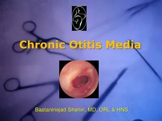
Definition • COM: unresolved inflammatory process of the middle ear and mastoid associated with TM perforation, otorrhea and hearing loss.
Etiology • Unresolved middle ear infection. • Uncomplicated inflammatory process of the middle ear may evolve over time to produce persistent effusion and irreversible mucosal change • Fluid contains enzymes to alter the mucosal lining of the middle ear, it results in collapse or chronic perforation • Obstruction of narrow communication between the antrum and the attic, the aditus.
Etiology • Dysfunction of Eustachian tube • Chronic inflammation in nose and pharynx • Dysfunction of immune system
Bacteriology • Pseudomonas aeruginosa (40-60%) • Straphylococus aureus (10-20%) • Anaerobic bacteria
Pathology • Middle ear mucosa is lined by secretory epithelium forming glandlike structure. • Hyalinization or tympanosclerosis • A healing response • It occurs during quiescent periods • It is formed by fused collagenous fibers • It is hardened by the deposition of calcium and phosphate crystals • Conductive hearing loss is associated with masses restricting ossicular mobility
Pathology • Ossicular erosion is frequent in COM • Infection process per se • Necrosis following vascular thrombosis • It most commonly affect the lenticular process of the incus and head of the stapes
Pathology • Cholesterol granulomas • Presence of yellowish masses surrounded by granulation tissue, edematous mucosa and fibrous tissue • It contains many cholesterol crystals and foreign body giant cells.
Pathology • Cholesteatoma: cystlike, expanding lesions of the temporal bone, lined by stratified epithelium and containing desquamated keratin and purulent material. • Classification • Congenital cholesteatoma • Acquired cholesteatoma
Pathology • Mechanics of mucosal transformation and epithelial ingrowth have been the focal point of cholesteatoma • Pocket retraction: dysfunction of Eustachian tube
Pathology • Epithelial migration: the edge of a peripheral perforation • Inward growth of the surface epithelium follows papillary proliferation of the germinative layer of the pars flaccida. • Metaplasia from pseudostratified ciliated columnar epithelium
Pathology • Pathogenesis of congenital cholesteatoma: • Ectodermal epithelial in proximity of the geniculate ganglion, medial to the neck of the malleus
Pathology • Diagnosis criteria: • Patients without previous history of ear disease, with normal and intact TM • The temporal bone pneumatization should be normal
Clinical presentations • Otorrhea • Malodorous associated with cholesteatoma • Hearing loss • Air conduction threshold is within 40 dB means TM proferation with intact ossicular chain • If air-bone gap is more than 40 dB is associated with discontinuity of ossicular chain
Clinical presentations • Physical findings • Defect in the pars tensa of TM or the pars flaccida or both • Atelectatic lesions in tensa or flaccida pars • Squamous epithelial invasion may invade middle ear • Granumoms, polyps, tympanosclerotic plaques in middle ear
Radiographic evaluation • Indications for image study • Uncontrollable aural discharge • Complications such as facial paralysis, labyrinthitis • When central nervous stystem involvement is suspected, MRI should be considered. • Coronal CT scan is perferred
Differential diagnosis • Tuberculous otitis media • Hematogenous route • Multiple perforation and fetid • Creamy aural discharge • Active pulmonary disease • Multiple antituberculosis agents
Differential diagnosis • Middle ear carcinoma • Middle age patient • Long term otorrhea with blood • Otalgia • Neoplasm in tympanum • CT scan showed temporal bone destruction
Managements • Medical treatment • Goals • Infection control • Stabilization of process • Prevention of irreversible damage and development of serious complications • 3%H202 clears up pus then antibiotics ear drops is used. • With the decrease of pus, 3% boric glycerin, 3% boric alcohol can be used • No aminoglycoside ear drops • No powders containing antibiotic and erosion ear drugs
Managements • Surgery • Goals • Safe ear: lesion removal • Dry ear • Hearing ear: reconstruction of ossiclar chain • classification • Myrigoplasty • Tympanoplasty • Tympanoplasty with mastoidectomy